. 2008 Mar 4;105(9):3545-50.
doi: 10.1073/pnas.0712275105. Epub 2008 Feb 22.

Overexpression of the dual-specificity phosphatase MKP-4/DUSP-9 protects against stress-induced insulin resistance

Affiliations

Overexpression of the dual-specificity phosphatase MKP-4/DUSP-9 protects against stress-induced insulin resistance

Brice Emanuelli et al. Proc Natl Acad Sci U S A. .

Abstract

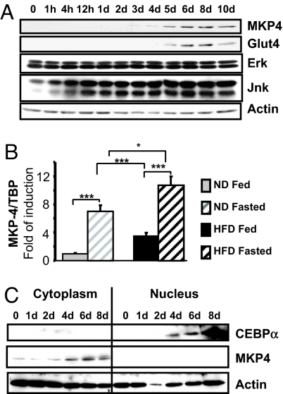
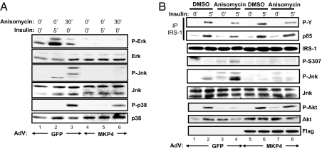
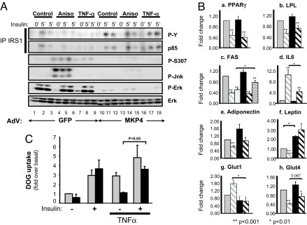
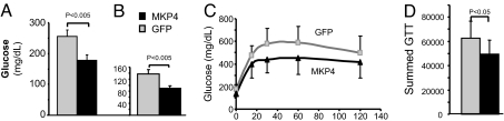
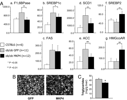
Insulin resistance, a hallmark of type 2 diabetes and obesity, is associated with increased activity of MAP and stress-activated protein (SAP) kinases, which results in decreased insulin signaling. Our goal was to investigate the role of MAP kinase phosphatase-4 (MKP-4) in modulating this process. We found that MKP-4 expression is up-regulated during adipocyte and myocyte differentiation in vitro and up-regulated during fasting in white adipose tissue in vivo. Overexpression of MKP-4 in 3T3-L1 cells inhibited ERK and JNK phosphorylation and, to a lesser extent, p38MAPK phosphorylation. As a result, the phosphorylation of IRS-1 serine 307 induced by anisomycin was abolished, leading to a sensitization of insulin signaling with recovery of insulin-stimulated IRS-1 tyrosine phosphorylation, IRS-1 docking with phosphatidylinositol 3-kinase, and Akt phosphorylation. MKP-4 also reversed the effect of TNF-alpha to inhibit insulin signaling; alter IL-6, Glut1 and Glut4 expression; and inhibit insulin-stimulated glucose uptake in 3T3-L1 adipocytes. Overexpression of MKP-4 in the liver of ob/ob mice decreased ERK and JNK phosphorylation, leading to a reduction in fed and fasted glycemia, improved glucose intolerance, decreased expression of gluconeogenic and lipogenic genes, and reduced hepatic steatosis. Thus, MKP-4 has a protective effect against the development of insulin resistance through its ability to dephosphorylate and inactivate crucial mediators of stress-induced insulin resistance, such as ERK and JNK, and increasing MKP-4 activity might provide a therapy for insulin-resistant disorders.


Conflict of interest statement

The authors declare no conflict of interest.

Figures

Fig. 1.
Fig. 1.
MKP-4 protein expression and localization in insulin-sensitive cells and tissues. (A) As specified in Experimental Methods, 3T3-L1 cells were induced to differentiate. Cell lysates were prepared at different time points after induction of differentiation and analyzed by immunoblots. Typical autoradiographs from at least three independent experiments are shown. (B) Mice were fed with either regular chow (10% fat, gray bars) or a high-fat diet (60% fat, black bars) for 11 weeks and then killed either in the early morning in the fed state (hatched bars) or after an overnight fast (gray and black bars). Expression of MKP-4 in epididymal adipose tissue was assessed by real-time PCR, normalized to TBP, and presented as the mean ± SEM. n ≥ 8 mice for each group. *, P < 0.05; **, P < 0.005; ***, P < 0.001. (C) Nuclear and cytoplasmic extracts were prepared at various times after induction of differentiation in 3T3-L1 cells and immunoblotted as specified. The first six lanes correspond to the cytoplasmic extracts, and the last six lanes correspond to the nuclear extracts.
Fig. 2.
Fig. 2.
MKP-4 inhibits ERK and JNK more than p38MAPK and protects against anisomycin-induced insulin resistance. The 3T3-L1ΔCAR cells were infected with equivalent amounts of AdV-GFP or AdV-MKP-4. (A) Forty-eight hours after infection, some wells were treated with anisomycin (5 μg/ml, 30 min) or with insulin (10 nM, 5 min). Levels of expression of total and serine-phosphorylated ERK, JNK, and p38MAPK were assessed by immunoblot analysis. (B) Forty-eight hours after infection, some wells were treated with anisomycin (5 μg/ml, 30 min) and then with insulin (10 nM, 5 min). Proteins from cell lysates were immunoprecipitated by using anti-IRS-1 antibodies, resolved by SDS/PAGE, and immunoblotted with anti-phosphotyrosine and anti-p85 antibodies or directly resolved by SDS/PAGE and then immunoblotted with antibodies as indicated.
Fig. 3.
Fig. 3.
MKP-4 protects against TNF-α-induced inhibition of insulin signaling and changes on gene expression. Differentiated 3T3-L1ΔCAR adipocytes were infected with equivalent amounts of control AdV-GFP or AdV-MKP-4. (A) Some wells were subsequently pretreated with anisomycin (5 μg/ml, 30 min) (lanes 4–6 and 13–15) or with TNF-α (25 ng/ml, 5 h) (lanes 7–9 and 16–18) and then stimulated with insulin (10 nM, 5 min). Proteins from cell lysates were immunoprecipitated by using anti-IRS-1 antibodies, resolved by SDS/PAGE, and immunoblotted with anti-phosphotyrosine and anti-p85 antibodies or directly resolved by SDS/PAGE and then immunoblotted with antibodies as indicated. (B) The 3T3-L1 adipocytes infected with AdV-GFP (gray bars) or AdV-MKP-4 (black bars) were treated (hatched bars) or not (gray and black bars) with TNF-α (25 ng/ml, 24 h) and with insulin (10 nM) during the last 8 h (dotted bars). mRNA expression was assessed by using real-time quantitative PCR in three independent experiments. Results are expressed as fold of stimulation and presented as the means ± SEM. (C) For 24 h before insulin stimulation (10 min at 100 nM), 3T3-L1 adipocytes infected with AdV-GFP (gray bars) or AdV-MKP-4 (black bars) were pretreated or not with TNF-α. At 5 min after insulin stimulation, 2-deoxyglucose uptake was measured. Means ± SEM of three independent experiments are shown.
Fig. 4.
Fig. 4.
MKP-4 overexpression reduces hyperglycemia and improves glucose intolerance in ob/ob mice. Ten-week-old ob/ob mice were infected with AdV-GFP (n = 5, gray) or AdV-MKP-4 (n = 6, black) via tail vein injection. (A and B) Fed (A) and fasting (B) blood glucose levels were measured 2 days after adenovirus injection. (C) An i.p. glucose tolerance test (GTT) was performed on day 5 after a 6-h fast. (D) The area under the curve was estimated by summing the numerical integration values of successive linear segments of the glucose curve for 0–15, 15–30, 30–60, and 60–120 min.
Fig. 5.
Fig. 5.
Effects of MKP-4 on gene expression and lipid accumulation in liver of ob/ob mice. (A) Gene-expression analysis was performed on liver extracts from 10-week-old control C57BL6 mice (n = 6, white bars) and ob/ob mice infected with AdV-GFP (n = 11, gray bars) or AdV-MKP-4 (n = 12, black bars). Hepatic expression of various genes was analyzed by real-time PCR. Expression levels were normalized to TBP and presented as the mean ± SEM. *, P < 0.05; **, P < 0.01. (B) Hematoxylin and eosin staining of liver section is shown at magnification ×40. (C) The triglyceride content in the liver of mice infected with AdV-GFP (n = 5, gray bars) or AdV-MKP-4 (n = 6, black bars) was measured as specified in Experimental Methods and presented as the means ± SEM. *, P < 0.05.

References

    1. Wellen KE, Hotamisligil GS. Inflammation, stress, and diabetes. J Clin Invest. 2005;115:1111–1119. - PMC - PubMed
    1. Engelman JA, Berg AH, Lewis RY, Lisanti MP, Scherer PE. Tumor necrosis factor alpha-mediated insulin resistance, but not dedifferentiation, is abrogated by MEK1/2 inhibitors in 3T3–L1 adipocytes. Mol Endocrinol. 2000;14:1557–1569. - PubMed
    1. Aguirre V, Uchida T, Yenush L, Davis R, White MF. The c-Jun NH (2)-terminal kinase promotes insulin resistance during association with insulin receptor substrate-1 and phosphorylation of Ser(307). J Biol Chem. 2000;275:9047–9054. - PubMed
    1. Hirosumi J, et al. A central role for JNK in obesity and insulin resistance. Nature. 2002;420:333–336. - PubMed
    1. Fujishiro M, et al. Three mitogen-activated protein kinases inhibit insulin signaling by different mechanisms in 3T3–L1 adipocytes. Mol Endocrinol. 2003;17:487–497. - PubMed

Publication types