. 2008 Mar 4;105(9):3449-54.
doi: 10.1073/pnas.0712126105. Epub 2008 Feb 22.

Genetic interaction between members of the Vangl family causes neural tube defects in mice

Affiliations

Genetic interaction between members of the Vangl family causes neural tube defects in mice

Elena Torban et al. Proc Natl Acad Sci U S A. .

Abstract

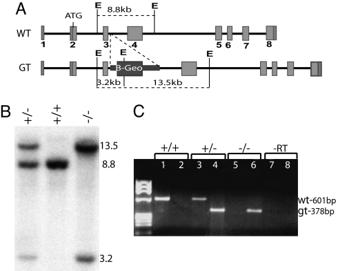
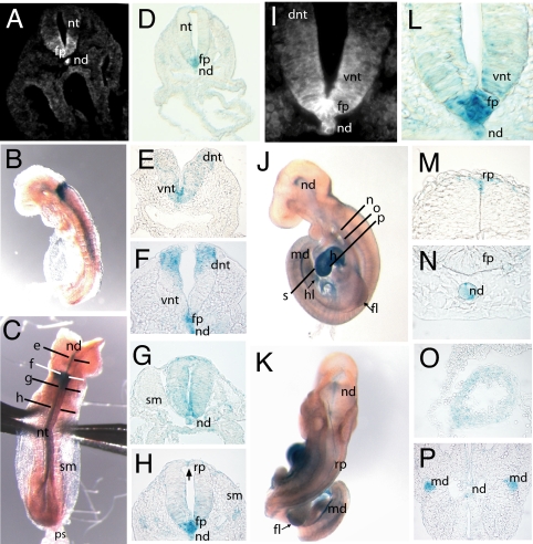
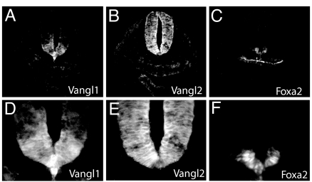
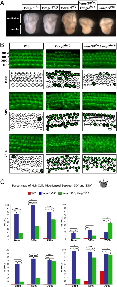
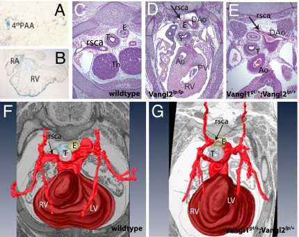
Neural tube defects (NTDs) are very frequent congenital abnormalities in humans. Recently, we have documented independent association of Vangl1 and Vangl2 gene mutations with NTDs. In the Looptail mouse, homozygosity (but not heterozygosity) for loss-of-function alleles at Vangl2 causes the severe NTD craniorachischisis, whereas heterozygosity for mutant variants of VANGL1 is associated with NTDs in a human cohort of sporadic and familial cases. To understand the role of Vangl1 in normal development, we created a mouse mutant with an inactivating mutation at Vangl1 (Vangl1(gt)). Vangl1 shows a dynamic pattern of expression in the developing neural tube and notochord at the time of neural tube closure. Vangl1(gt/+) heterozygotes and Vangl1(gt/gt) homozygotes are viable and fertile, although Vangl1(gt/gt) display subtle alterations in polarity of inner hair cells of the cochlea. Remarkably, and as opposed to healthy Vangl1(gt/+) and Vangl2(lp/+) heterozygotes, Vangl1(gt/+);Vangl2(lp/+) double heterozygotes show profound developmental defects that include severe craniorachischisis, inner ear defects (disorganization of the stereociliary bundles of hair cells of the organ of Corti), and cardiac abnormality (aberrant right subclavian artery). These results show that genetic interaction between Vangl1 and Vangl2 genes causes neural tube defects and raise the possibility that interaction between individual Vangl genes and other genetic loci and/or environmental factors may additionally contribute to the etiology of NTDs.


Conflict of interest statement

The authors declare no conflict of interest.

Figures

Fig. 1.
Fig. 1.
Generation of the Vangl1 mutant transgenic mouse line. (A) Schematic representation of WT and targeted Vangl1 alleles, including the inserted β-gal–Neomycin resistance gene-trap cassette. (B) WT and targeted Vangl1 alleles distinguished by Southern blotting of genomic DNA digested with EcoRV (E) and probed with a 601-bp exon 3–4 cDNA probe, which yields fragments of 8.8 kb (WT) vs. 3.2/13.5 kb (targeted). (C) Diagnostic RT-PCR to amplify WT (601 bp; lanes 1, 2, 5, and 7) and mutant (378 bp; lanes 2, 4, 6, and 8) Vangl1 transcripts.
Fig. 2.
Fig. 2.
Vangl1 expression during neurulation. (A and I) Detection of Vangl1 protein in E8.5 WT embryos by using a rabbit anti-Vangl1 polyclonal antiserum (magnification, ×200 and ×630). (B and C) Whole-mount X-gal staining on E8.5 Vangl1gt/gt embryos showing lateral and dorsal views, respectively. The section planes used for E–H are shown in C. (D and L) Section of Vangl1gt/gt E8.5 embryo stained with X-gal showing a pattern of Vangl1 expression in notochord and floor plate very similar to that detected with anti-Vangl1 antiserum (magnification, ×200 and ×630) (E–H). Transverse sections and X-gal staining of neural tube (E8.5) arranged in rostral–caudal progression (magnification, ×400). (J and K) Whole-mount X-gal staining of Vangl1gt/gt E9.5 embryos; the section planes used for M–P are shown in K. (M–P) X-Gal staining of E9.5 Vangl1gt/gt embryos; transverse sections (magnification, ×400). (O and P) X-Gal staining of myocardial cells of heart (O) and epithelium of mesonephric duct (P). fp, floor plate; dnt, dorsal neural tube; h, heart; hl, hindlimb; md, mesonephric duct; nd, notochord; nt, neural tube; ps, primitive streak; rp, roof plate; sm, somites; vnt, ventral neural tube.
Fig. 3.
Fig. 3.
Vangl1 and Vangl2 protein coexpression in neural tube. Serial transverse sections of E8.5 embryos stained with anti-Vangl1 (A and D), anti-Vangl2 (B and E), and anti-Foxa2 (C and F) antisera. Magnification, ×200 (A–C ); ×630 (D–F ).
Fig. 4.
Fig. 4.
Genetic interaction between Vangl1 and Vangl2 during neural tube closure. The presence of craniorachischisis in Vangl2lp/lp (E) and in Vangl1gt/+;Vangl2lp/+ double heterozygote embryos (arrows in C and D) is compared with phenotypically normal Vangl1gt/+;Vangl2+/+ controls (A and B). E13.5 embryos are shown in A–D. E18.5 embryos are shown in E and F. B and D are dorsal views of embryos in A and C. Closed vs. open eyelid is identified by white arrows in E and F.
Fig. 5.
Fig. 5.
Genetic interaction of Vangl1 and Vangl2 in planar cell polarity of the neurosensory cells in the inner ear. (A) Macroanatomy of inner ear structures dissected from E18.5 embryos of various Vangl1 and Vangl2 genotypes is shown. (B) Whole-mount preparations of organs of Corti from E18.5 WT, Vangl2lp/lp, and Vangl1gt/+;Vangl2lp/+ embryos stained with phalloidin–FITC antibody to visualize actin-based stereociliary bundles. The analyzed regions were determined relative to the length of the cochlear duct. The base region was 5% from the most basal position of the cochlea, the 50% region was the midpoint between the most basal and most apical ends, and the apical region was at 75% from the most basal position. Each microscopic image is schematically depicted: White circles correspond to the hair cells with normal planar orientation of the stereociliary bundles, and the black circles correspond to the hair cells with randomized orientation of the actin vertexes. The individual layers of IHCs and OHCs (OHC1–3) are identified. (C) Quantification of the number of misoriented hair cells (as a percentage) found in embryos of different Vangl1 and Vangl2 genotypes. The asterisks indicate the level of significance (χ2 analysis): ***, P < 0.0005; **, P ≥ 0.0005 to P < 0.005; *, P ≥ 0.005 to P < 0.05. The absence of an asterisk indicates lack of significance (P ≥ 0.05).
Fig. 6.
Fig. 6.
Vangl1gt/+;Vangl2lp/+ embryos exhibit aberrant right subclavian artery. (A) Vangl1 expression in fourth aortic arch of E10.5 Vangl1gt/gt embryo (X-gal staining; magnification, ×600). (B) X-Gal staining of the intracardiac structures of the of E10.5 Vangl1gt/gt embryos (magnification, ×200). (C and D) Transverse sections through the hearts of E14.5 embryos (magnification, ×400). (C) In WT embryos, the right subclavian artery (arrows) is positioned laterally from trachea and esophagus. In Vangl2lp/lp embryos (D) and in Vangl1gt/+;Vangl2lp/+ (E), the right subclavian artery is positioned dorsal to esophagus. (F and G) Three-dimensional reconstruction of the arteries of E14.5 WT (F) and Vangl1gt/+;Vangl2lp/+ hearts (G). Ao, aorta; DAo, dorsal aorta; E, esophagus; LV, left ventricle; PAA, pharyngeal arch artery; PV, pulmonary valve; RA, right atrium; rsca, right subclavian artery; RV, right ventricle; T, trachea; Th, thymic primordium.

References

    1. Frey L, Hauser WA. Epidemiology of neural tube defects. Epilepsia. 2003;44(Suppl)(3):4–13. - PubMed
    1. Botto LD, Moore CA, Khoury MJ, Erickson JD. Neural-tube defects. N Engl J Med. 1999;341:1509–1519. - PubMed
    1. Kibar Z, Capra V, Gros P. Toward understanding the genetic basis of neural tube defects. Clin Genet. 2007;71:295–310. - PubMed
    1. Finnell RH, Waes JG, Eudy JD, Rosenquist TH. Molecular basis of environmentally induced birth defects. Annu Rev Pharmacol Toxicol. 2002;42:181–208. - PubMed
    1. Copp AJ, Greene ND, Murdoch JN. The genetic basis of mammalian neurulation. Nat Rev Genet. 2003;4:784–793. - PubMed

Publication types