. 2008 Mar 4;105(9):3232-7.
doi: 10.1073/pnas.0710412105. Epub 2008 Feb 22.

Engineering of mucin-type human glycoproteins in yeast cells

Affiliations

Engineering of mucin-type human glycoproteins in yeast cells

Koh Amano et al. Proc Natl Acad Sci U S A. .

Abstract

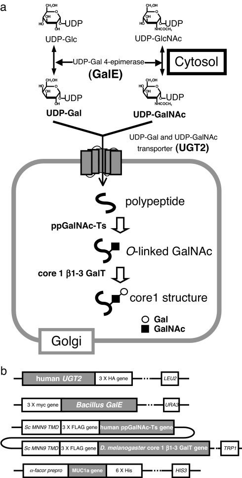
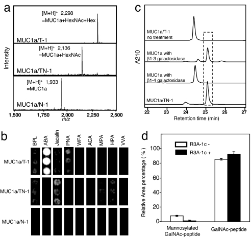
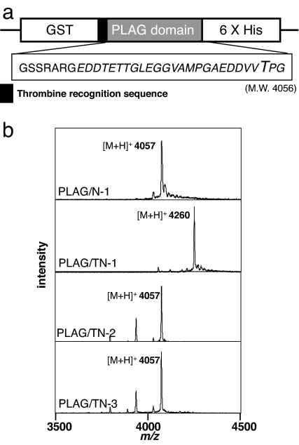
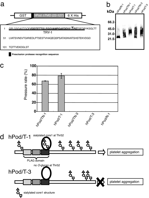
Mucin-type O-glycans are the most typical O-glycans found in mammalian cells and assume many different biological roles. Here, we report a genetic engineered yeast strain capable of producing mucin-type sugar chains. Genes encoding Bacillus subtilis UDP-Gal/GalNAc 4-epimerase, human UDP-Gal/GalNAc transporter, human ppGalNAc-T1, and Drosophila melanogaster core1 beta1-3 GalT were introduced into Saccharomyces cerevisiae. The engineered yeast was able to produce a MUC1a peptide containing O-glycan and also a mucin-like glycoprotein, human podoplanin (hPod; also known as aggrus), which is a platelet-aggregating factor that requires a sialyl-core1 structure for activity. After in vitro sialylation, hPod from yeast could induce platelet aggregation. Interestingly, substitution of ppGalNAc-T1 for ppGalNAc-T3 caused a loss of platelet aggregation-inducing activity, despite the fact that the sialyl-core1 was detectable in both hPod proteins on a lectin microarray. Most of O-mannosylation, a common modification in yeast, to MUC1a was suppressed by the addition of a rhodanine-3-acetic acid derivative in the culture medium. The yeast system we describe here is able to produce glycoproteins modified at different glycosylation sites and has the potential for use in basic research and pharmaceutical applications.


Conflict of interest statement

The authors declare no conflict of interest.

Figures

Fig. 1.
Fig. 1.
Engineering of a mucin-type glycosylation pathway in yeast. (a) Introduction of a mucin-type glycosylation pathway in yeast. UDP-Gal and UDP-GalNAc are synthesized from UDP-Glc and UDP-GlcNAc, respectively, by GalE protein in the cytosol. UGT2 protein transports UDP-Gal and UDP-GalNAc from the cytosol to the Golgi lumen. Next, ppGalNAc-Ts and core1 β1–3 GalT transfer GalNAc and Gal to polypeptides. (b) Construction of each gene integrated into the yeast genome. UGT2 protein was C-terminally 3× HA-tagged, and GalE protein had an N-terminal 3× myc tag. The glycosyltransferases were fused to the transmembrane domain of yeast MNN9, and a 3× FLAG-tag was inserted between the transmembrane domain and the catalytic domains of the transferases. MUC1a was fused to the α-factor prepro sequence for secretion into the medium, and a hexa-histidine tag was added to the C terminus.
Fig. 2.
Fig. 2.
Analysis of MUC1a peptide expressed in engineered yeast cells. (a) MALDI-TOF MS spectra of each exogenously expressed MUC1a peptide. MUC1a produced by N-1, TN-1, and T-1 cells show MUC1a/N-1, MUC1a/TN-1, and MUC1a/T-1, respectively. The molecular mass of MUC1a is 1,932. (b) Lectin microarray. BPL, Bauhinia purpurea lectin; ABA, Agaricus bisporus agglutinin; Jacalin, Artocarpus integrifolia lectin; PNA, peanut agglutinin; WFA, Wisteria floribunda agglutinin; ACA, Amaranthus caudatus agglutinin; MPA, Maclura pomifera agglutinin; HPA, Helix pomatia agglutinin; VVA, Vicia villosa agglutinin. GalNAc binders: Jacalin, WFA, MPA, HPA, and VVA; core1 binders: ABA, PNA, and ACA; core1 and GalNAc binder: BPL. (c) Enzymatic digestion of MUC1a/T-1. The peak at 24.5 min corresponds to a core1-MUC1a peptide, whereas the peak at 25.1 min corresponds to the GalNAc-MUC1a peptide. (d) Inhibition of yeast O-mannosylation with a chemical reagent. MUC1a was expressed in TN-1 strains in the presence or absence of R3A-1c reagent. The total area of the recombinant MUCla peak on the chromatogram is set to 100%, and error bars show standard deviation of the mean (n = 3).
Fig. 3.
Fig. 3.
ppGalNAc-T1 transfers GalNAc to the PLAG peptide. (a) PLAG peptide was expressed with an N-terminal GST tag and a C-terminal hexa-histidine tag. The construct contains a thrombin recognition sequence (LVPRGS) between GST and the PLAG-peptide coding region. MALDI-TOF MS analysis of the PLAG peptide was done after thrombin digestion. The amino acid sequence of PLAG peptide after thrombin digestion is GSSRARGEDDTETTGLEGGVAMPGAEDDVVTPG. (b) The molecular mass of the PLAG peptide is 4,056. PLAG/N-1, PLAG/TN-1, PLAG/TN-2, and PLAG/TN-3 refer to PLAG peptide produced by the N-1, TN-1, TN-2, and TN-3 yeast strains, respectively.
Fig. 4.
Fig. 4.
Expression and analysis of PLAG domain and human podoplanin (hPod). (a) A construct that expresses hPod with an N-terminal GST tag and a C-terminal hexa-histidine tag. The resultant recombinant protein contains a PreScission Protease recognition sequence (LEVLFQGP) between GST and hPod. The amino acid sequence of the TRY-1 trypsin fragment is double-underlined, and the PLAG domain is in italics. Big T, the essential Thr residue for platelet-aggregation inducing activity. (b) Immunoblot analysis of recombinant hPod. hPod/TN-1, hPod/T-1, hPod/TN-3, hPod/T-3, and hPod/N-1 shows hPod produced by TN-1 cells, T-1 cells, TN-3 cells, T-3 cells, and N-1 cells. (c) Platelet aggregation-inducing activity of recombinant hPod after in vitro sialylation. Before sialylation, no sample induced platelet aggregation (data not shown). Platelet aggregation was measured using WBA carna with the screen-filtration method. Pressure means a value of a pressure required for sample to pass through a filter. The pressure rate that a sample cannot pass through the filter shows 100%. The normalized mean ± SD of three independent experiments is shown. (d) Both hPod/T-1 and hPod/T-3 are modified with mucin-type O-glycan, although they are glycosylated at different sites. Thus, hPod/T-1, but not hPod/T-3, contains sialylated mucin-type O-glycan at Thr-52, and hPod/T-1 can induce platelet aggregation whereas hPod/T-3 cannot. All O-glycosylation sites of podoplanin except at Thr-52 are estimated O-glycosylation sites.

References

    1. Birch NP, Estivariz FE, Bennett HP, Loh YP. Differential glycosylation of N-POMC1–77 regulates the production of gamma 3-MSH by purified pro-opiomelanocortin converting enzyme. FEBS Lett. 1991;290:191–194. - PubMed
    1. Atiya-Nasagi Y, Cohen H, Medalia O, Fukudan M, Sagi-Eisenberg R. O-glycosylation is essential for intracellular targeting of synaptotagmins I, II in non-neuronal specialized secretory cells. J Cell Sci. 2005;118:1363–1372. - PubMed
    1. Tian E, Ten Hagen KG. A UDP-GalNAc:polypeptide N-acetylgalactosaminyltransferase is required for epithelial tube formation. J Biol Chem. 2007;282:606–614. - PubMed
    1. Gendler SJ, Spicer AP. Epithelial mucin genes. Annu Rev Physiol. 1995;57:607–634. - PubMed
    1. Perez-Vilar J, Hill RL. The structure and assembly of secreted mucins. J Biol Chem. 1999;274:31751–31754. - PubMed

Publication types

LinkOut - more resources