. 2008 Jul;7(7):1378-88.
doi: 10.1074/mcp.M800069-MCP200. Epub 2008 Feb 22.

Identification of CKAP4/p63 as a major substrate of the palmitoyl acyltransferase DHHC2, a putative tumor suppressor, using a novel proteomics method

Affiliations

Identification of CKAP4/p63 as a major substrate of the palmitoyl acyltransferase DHHC2, a putative tumor suppressor, using a novel proteomics method

Jun Zhang et al. Mol Cell Proteomics. 2008 Jul.

Abstract

Protein palmitoylation is the post-translational addition of the 16-carbon fatty acid palmitate to specific cysteine residues by a labile thioester linkage. Palmitoylation is mediated by a family of at least 23 palmitoyl acyltransferases (PATs) characterized by an Asp-His-His-Cys (DHHC) motif. Many palmitoylated proteins have been identified, but PAT-substrate relationships are mostly unknown. Here we present a method called palmitoyl-cysteine isolation capture and analysis (or PICA) to identify PAT-substrate relationships in a living vertebrate system and demonstrate its effectiveness by identifying CKAP4/p63 as a substrate of DHHC2, a putative tumor suppressor.


Figures
F<sc>ig</sc>. 1.
Fig. 1.
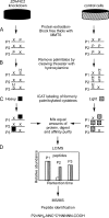
PICA: determining PAT-substrate specificity by differential labeling of palmitoylated proteins with ICAT. A, in one set of cultures, ZDHHC2 expression is knocked down by transfecting HeLa cells with ZDHHC2-specific siRNA (Dharmacon). Proteins are extracted from experimental and control cells and treated with the thiol-specific blocking reagent MMTS. This step chemically modifies or protects all thiols (“X” on the proteins P1–P3) that are free at physiological pH and leaves the palmitoylated cysteines (P) undisturbed as depicted on P1–P3. B, following the protection or blocking of free thiols, palmitates are removed by selective cleavage of the thioester bond with hydroxylamine at pH 7.4, which generates a distinctive set of free, formerly palmitoylated, reactive thiols (C) that can be selectively labeled with ICAT reagents. The iodoacetamide moiety at one end of the ICAT reagent reacts with the thiol side chain of cysteines; on the other end, biotin provides a mechanism for affinity purification of thiol-captured peptides on an avidin column. D, proteins from knockdown and control conditions are mixed in equal amounts and digested in-gel with trypsin. ICAT-labeled peptides are enriched by avidin affinity purification and analyzed by LC-MS/MS. A pair of ICAT-labeled peptides is chemically identical and is easily visualized because they essentially coelute. The different isotopic composition of the H and L ICATs provides a 9-Da difference in mass that is easily measured by mass spectrometry. Even if equal amounts of a single protein exist in two different samples, the quantity of protein that is captured depends directly on its degree of palmitoylation: if all of a single protein is palmitoylated under one condition, then all of it will be captured; if only half of this protein is palmitoylated under another condition, then the capture rate of that protein will be half as much, relative to control, making it appear half as abundant. Peptide abundance values derived from the areas of commonly shared isotopic peaks were calculated by Analyst/BioAnalyst (Applied Biosystems). Proteins for which there has been no change in palmitoylation (i.e. equal capture rates) will yield an H/L ratio of 1. The degree to which palmitoylation is diminished will register as a decrease in the H/L ratio (i.e. 50% reduction in palmitoylation will correspond to an H/L ratio of 0.5). A change in the capture rate that results in a change in the postpurification abundance is measured in the LC-MS phase. E, finally the peptides are further analyzed via MS/MS, which permits identification of the proteins corresponding to the captured peptides after searching the tandem MS data against a protein sequence database.
F<sc>ig</sc>. 2.
Fig. 2.
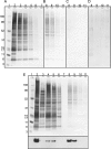
The specificity of PICA for palmitoylation. In each panel, captured proteins are separated by SDS-PAGE and silver-stained. 20 μl of a 100-μl wash or elution was loaded into each lane. A, following the capture of proteins on the thiol affinity column, nonspecifically bound proteins were removed by a series of stringent washes. Lane 1 is the relative mobility markers; lane 2 is the whole protein following MMTS block; lane 3 is the flow-through after overnight protein binding. The last of the washes in the series is shown in A (lanes 4–7) to illustrate that many proteins are removed in the initial washes (lanes 4–6) (1% SDS in 50 mm Tris buffer (pH 7.4)) and that by wash 20 (lane 7) proteins are no longer detected in the washes. B, lanes 8–11 contain a select set of putative formerly palmitoylated proteins eluted from the column after successive washes in elution buffer (1% SDS, 50 mm Tris buffer (pH 7.4), 100 μm β-ME) that are putatively palmitoylated. C, PICA fails to capture proteins (lanes 8–11, eluted as in B) when there is no hydroxylamine treatment after blocking free thiols with MMTS illustrating that MMTS is blocking quantitatively and that the thioester bond linking palmitates to cysteines is also stable under these experimental conditions. D, to test further the specificity of PICA for palmitoylation, we repeated the experiment using HeLa cells that had been treated for 6 h with 100 μm 2BP, an inhibitor of palmitoylation. This treatment eliminated capture of proteins (lanes 8–11, eluted as in B and C). Blocking palmitoylation with 2BP leaves normally palmitoylated cysteines unmodified by palmitate and available for modification by MMTS, thereby eliminating their capture in subsequent steps. E, silver-stained SDS-PAGE gel and Western blot of proteins captured by PICA from MDCK cells stably expressing GYH. Lanes 1–7 are the same conditions as lanes 1–7 in A. Lanes 8–11 are the same elution conditions as in B, C, and D. Western blot of GYH from the corresponding samples shows that capture of GYH is not quantitative as some appears in washes 4 and 5, and the majority of GYH is released during elution (lanes 8 and 9). The membrane was probed with anti-GFP primary antibody (Clontech) followed by horseradish peroxidase-conjugated secondary antibody (Pierce). Detection was by enhanced chemiluminescence (Pierce); the exposure time was 1 min.
F<sc>ig</sc>. 3.
Fig. 3.
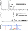
Mass spectrometry identification of CKAP4/p63 peptides labeled by H and L ICAT reagents. A, ICAT-labeled tryptic peptides (m/z 753.9 and 756.9) derived from a putative palmitoylated protein and detected by LC-MS/MS in the retention time range 46–49 min. Following a Mascot protein database search of the corresponding MS/MS spectrum, the sequence was determined to be SSSSSSASAAAAAAAASSSASC*SR, a tryptic peptide of CKAP/p63 modified at the cysteine residue with the ICAT reagent. The specificity of the labeling reaction allows for selective derivatization of palmitoyl-cysteines by the ICAT reagent. The 9-Da mass difference (Δm/z 3 for a triply charged species) provided by the heavy ICAT reagent can be observed for this particular ICAT pair, and any variation in mass spectral intensity correlates to changes in protein palmitoylation induced from the ZDHHC2 knockdown treatment. B, reconstructed ion chromatograms derived from the monoisotopic masses corresponding to the L and H ICAT-labeled peptide SSSSSSASAAAAAAAASSSASC*SR from a separate experiment. Upon calculation of the peak area ratios (H/L) obtained from four separate experiments using a quantitation algorithm within the data acquisition software, this particular peptide showed a 35% decrease in palmitoylation (p < 0.001, n = 4 complete independent PICA runs from cell culture to mass spectrometry runs) after ZDHHC2 knockdown. C, MS/MS spectrum of a triply charged ion at m/z 757.06. After a protein database search using Mascot, the peptide sequence was determined to be SSSSSSASAAAAAAAASSSASC*SR (Mascot ion score of 64.6), a heavy ICAT-labeled tryptic peptide of CKAP/p63. The presence of the isobaric fragment ions y1/b2 and y2/b3 (Δm/z 0.05) made unambiguous assignment difficult for these particular ions given the mass measurement accuracy specified for data processing and subsequent database searching. However, after inspection of the MS/MS raw data, adequate mass resolving power and peak shape were obtained to identify both y1 and b2 ions. The mass accuracy of the MS/MS spectrum allowed for putative assignment of the peak at m/z 262 as the y2 fragment ion, although the b3 fragment ion should not be precluded because of limited resolving power and peak shape obtained at this m/z value. Only one of the four data sets for the identification of CKAP4/p63 is shown here. It is representative of the other three. cps, counts/s.
F<sc>ig</sc>. 4.
Fig. 4.
Confirmation that CKAP4/p63 is a substrate of DHHC2. Co-overexpression of CKAP4/p63 and ZDHHC2 in COS cells resulted in a 5.7-fold increase in [3H]palmitate labeling of CKAP4/p63 (lane 4) versus control (lane 1) confirming that CKAP4/p63 is a substrate of DHHC2. Overexpression of ZDHHC2 alone (lane 2) or CKAP4/p63 alone (lane3) resulted in much smaller signal increases versus control. Treatment of the protein sample run in lane 4 with hydroxylamine removes [3H]palmitate from CKAP4/p63 confirming that [3H]palmitate is attached by a thioester bond (lane 5). Equal amounts of total protein lysate were added to each lane. The fluorographic signal was the result of a 24-h exposure. The integrated signal density of each band was measured using ImageJ (42) and normalized to the mock-transfected control, lane 1.
F<sc>ig</sc>. 5.
Fig. 5.
CKAP4/p63 localization is redistributed to perinuclear ER membranes from the PM when DHHC2-mediated palmitoylation is blocked by siRNA-mediated ZDHHC2 knockdown. A, CKAP4/p63 immunolocalization (TRITC channel) in untreated HeLa cells co-localizes with the PM marker, pan-cadherin (FITC channel). B, in HeLa cells treated for 48 h with siRNA targeting ZDHHC2, CKPA4/p63 immunostaining is no longer detected on the PM but is instead confined to endomembranes (ER) that closely surrounded the nucleus. The collapse of the ER around the nucleus in response to reduced palmitoylation of CKAP4/p63 is similar to what is seen when deletion of phosphomimicking mutants of CKAP4/p63, which are unable to bind microtubules, are expressed in cells (25).

References

    1. Roth, A. F., Wan, J., Bailey, A. O., Sun, B., Kuchar, J. A., Green, W. N., Phinney, B. S., Yates, J. R., III, and Davis, N. G. ( 2006) Global analysis of protein palmitoylation in yeast. Cell 125, 1003–1013 - PMC - PubMed
    1. Fukata, M., Fukata, Y., Adesnik, H., Nicoll, R. A., and Bredt, D. S. ( 2004) Identification of PSD-95 palmitoylating enzymes. Neuron 44, 987–996 - PubMed
    1. Drisdel, R. C., and Green, W. N. ( 2004) Labeling and quantifying sites of protein palmitoylation. BioTechniques 36, 276–285 - PubMed
    1. Oyama, T., Miyoshi, Y., Koyama, K., Nakagawa, H., Yamori, T., Ito, T., Matsuda, H., Arakawa, H., and Nakamura, Y. ( 2000) Isolation of a novel gene on 8p21.3-22 whose expression is reduced significantly in human colorectal cancers with liver metastasis. Genes Chromosomes Cancer 29, 9–15 - PubMed
    1. Wong, M. L., and Medrano, J. F. ( 2005) Real-time PCR for mRNA quantitation. BioTechniques 39, 75–85 - PubMed

Publication types