. 2008 Mar;26(3):326-34.
doi: 10.1038/nbt1390. Epub 2008 Feb 24.

Engineered lentivector targeting of dendritic cells for in vivo immunization

Affiliations

Engineered lentivector targeting of dendritic cells for in vivo immunization

Lili Yang et al. Nat Biotechnol. 2008 Mar.

Abstract

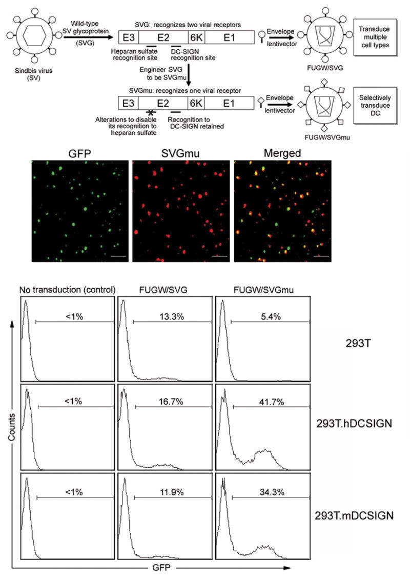
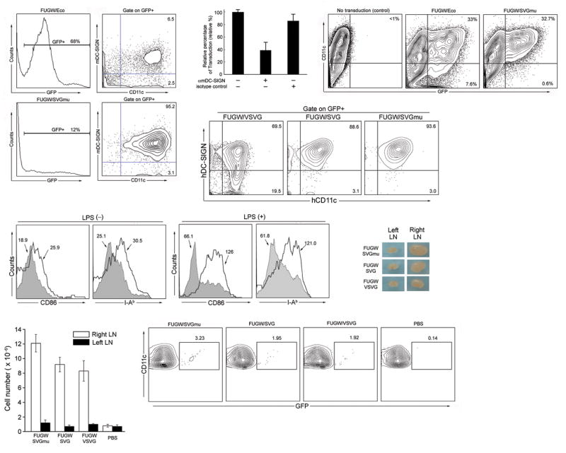
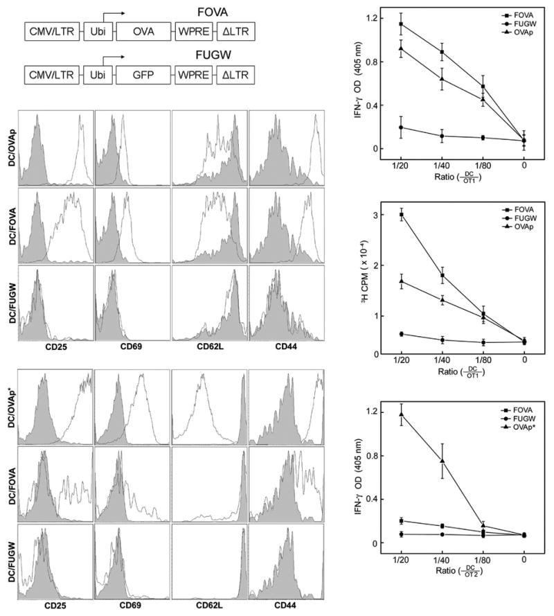
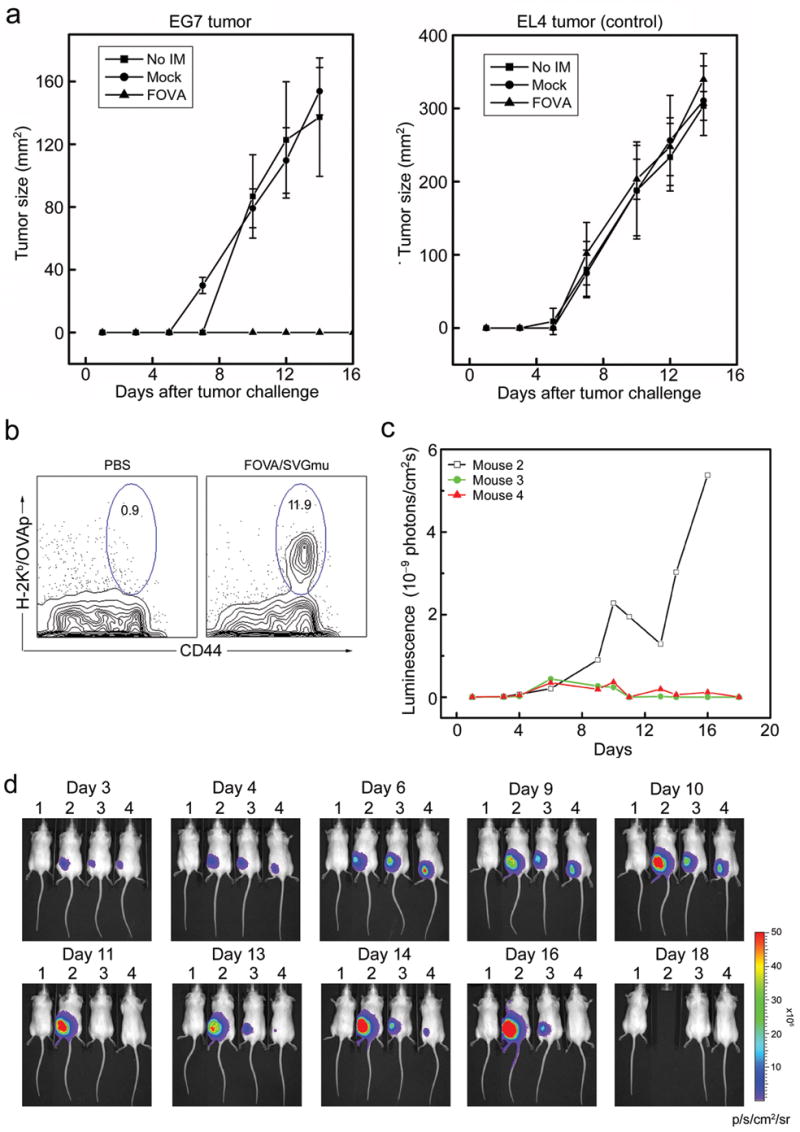
We report a method of inducing antigen production in dendritic cells by in vivo targeting with lentiviral vectors that specifically bind to the dendritic cell-surface protein DC-SIGN. To target dendritic cells, we enveloped the lentivector with a viral glycoprotein from Sindbis virus engineered to be DC-SIGN-specific. In vitro, this lentivector specifically transduced dendritic cells and induced dendritic cell maturation. A high frequency (up to 12%) of ovalbumin (OVA)-specific CD8(+) T cells and a significant antibody response were observed 2 weeks after injection of a targeted lentiviral vector encoding an OVA transgene into naive mice. This approach also protected against the growth of OVA-expressing E.G7 tumors and induced regression of established tumors. Thus, lentiviral vectors targeting dendritic cells provide a simple method of producing effective immunity and may provide an alternative route for immunization with protein antigens.


Conflict of interest statement

COMPETING INTERESTS STATEMENT: The authors declare that they have no competing financial interests.

Figures

Figure 1
Figure 1
Lentivector bearing engineered Sindbis viral glycoprotein targets to DC-SIGN-expressing cells. (a) A schematic representation of the general strategy to engineer a lentivector system capable of targeting DCs. (b) Viral supernatant harvested from virus-producing cells transiently transfected with GFP-vpr, SVGmu, and other necessary packaging constructs was coated to a poly-lysine containing coverslip by centrifugation. A hemagglutinin (HA) epitope tag (YPYDVPDYA) was engineered into SVGmu to facilitate its detection by antibody. The resulting coverslip was then rinsed and immunostained with an anti-HA tag antibody (red) to label SVGmu and imaged using a laser confocal microscope. The scale bar represents 2 μm. (c) One milliliter of fresh viral supernatants of FUGW/SVG and FUGW/SVGmu were used to transduce 293T cells (2×10⁵) expressing human DC-SIGN (293T.hDCSIGN) or murine DC-SIGN (293T.mDCSIGN). The parental 293T cells lacking the expression of DC-SIGN were included as controls. Three days later, the transduction efficiency was measured by analyzing GFP expression using flow cytometry. The specific transduction titer of FUGW/SVGmu was estimated to be ∼1×10⁶ TU/ml for 293T.hDC-SIGN and ∼0.8×10⁶ TU/ml for 293T.mDC-SIGN.
Figure 2
Figure 2
Lentivector encoding a reporter GFP gene and bearing SVGmu can selectively transduce DCs in vitro and in vivo. (a) Whole bone marrow cells isolated from B6 mice were exposed to the fresh viral supernatant of FUGW/SVGmu. The FUGW lentivector pseudotyped with the ecotropic glycoprotein (FUGW/Eco) was included as a non-targeting control. Three days post-transduction, the cells were collected for flow cytometric analysis of GFP expression. Surface antigens of the GFP-positive cells were assessed by staining with anti-CD11c and anti-DC-SIGN antibodies. (b) Anti-murine DC-SIGN antibody was added into viral supernatant during transduction of whole mouse bone marrow cells for 8 h. Then, the supernatant was replaced with fresh medium. The cells were analyzed for GFP expression after 2 days. Isotype-matched antibody was used as a control. (c) Murine bone marrow-derived DCs (mBMDCs) were generated by culturing freshly isolated bone marrow cells in the presence of cytokine GM-CSF for 6 days. The resulting cells were transduced with the fresh viral supernatant of either the targeting FUGW/SVGmu or non-targeting FUGW/Eco vector. GFP and CD11c expression were measured by flow cytometry. (d) Human monocyte-derived DCs (hMoDCs) were generated by culturing freshly purified CD14+ peripheral blood moncytes in the presence of GM-CSF and IL-4. The cells from the day 2 culture were transduced with the fresh viral supernatant of either FUGW/SVGmu, or FUGW/VSVG, or FUGW/SVG vector. GFP expression was measured by flow cytometry at day 6. (e) Upon targeted transduction of mouse BMDCs with FUGW/SVGmu, DC activation was assessed by analyzing the surface expression of CD86 and I-Ab using flow cytometry. The addition of LPS (1 μg/ml) overnight was used as a synergistic stimulator for the activation of transduced BMDCs. Shaded area, GFP negative cells (untransduced); solid line, GFP positive cells (transduced). (f-h) In vivo DC-targeting using FUGW/SVGmu lentivector. B6 mice were injected with 50×106 TU of FUGW/SVGmu, FUGW/SVG, or FUGW/VSVG, and analyzed 3 days later. Mice injected with PBS were included as a control. (f) Comparison of a representative inguinal lymph node close to the injection site (right) and the equivalent lymph node distant from the injection site (left). (g) Total cell number counts of the indicated lymph nodes. (h) Representative flow cytometric analysis of CD11c+ cells from the indicated lymph nodes that are close to the injection sites. The numbers indicate the fraction of GFP+ DC populations.
Figure 3
Figure 3
Mouse bone marrow-derived DCs (mBMDCs) transduced by a SVGmu enveloped lentivector encoding an OVA gene can stimulate OVA-specific CD8+ and CD4+ T cells in vitro. (a) A schematic representation of the lentivector encoding the OVA antigen (FOVA) or the lentivector encoding GFP (FUGW) as a control. LTR: long terminal repeat; Ubi: human ubiquitin-C promoter; WPRE: woodchuck hepatitis virus posttranscriptional regulatory element. (b-f) OVA-specific, CD8+ OT1 T cells and CD4+ OT2 T cells were harvested from the spleens of OT1 TCR- or OT2 TCR-transgenic mice (The Jackson Laboratory) and were cocultured with FOVA/SVGmu-transduced mBMDCs (DC/FOVA) in vitro for 3 days. Non-transduced BMDC pulsed with either OVAp peptide (SIINFEKL) (DC/OVAp), recognized by OT1 T cells, or OVAp* peptide (ISQAVHAAHAEINEAGR) (DC/OVAp*), recognized by OT2 T cells, were included as positive controls. mBMDCs transduced with FUGW/SVGmu (DC/FUGW) were included as a negative control. (b) Patterns of surface activation markers of OT1 T cells cocultured with various mBMDCs were assessed by antibody staining for CD25, CD69, CD62L, and CD44. Shaded area: naïve OT1 T cells harvested from transgenic animals; solid line: OT1 T cells cocultured with the indicated mBMDCs. (c) OT1 T cells were mixed with various dilutions of mBMDCs transduced with FOVA/SVGmu (■), FUGW/SVGmu (●), or pulsed with OVAp peptide (▲) and cultured for 3 days. Secretion of IFN-γ was measured by ELISA. (d) The proliferative responses of treated OT1 T cells from (c) were measured by a [3H] thymidine incorporation assay. (e) Similar to (b) except for the analysis of activated CD4+ OT2 T cells. Shaded area, naïve OT2 T cells harvested from transgenic animals; solid line, OT2 T cells cocultured with BMDCs. (f) Similar to (c) except for the responding cells being CD4+ OT2 T cells.
Figure 4
Figure 4
In vivo stimulation of antigen specific T cell and antibody responses in wild-type B6 mice following a subcutaneous injection of the DC-targeting lentivector FOVA/SVGmu. (a) B6 mice were immunized subcutaneously with 50×106 TU of either FOVA/SVGmu or FUGW/SVGmu (as a control). Mice without immunization (no imm.) were included as a negative control. Fourteen days post-immunization, spleen cells were harvested and analyzed for the presence of OVA-specific CD8+ T cells measured by H-2Kb-SIINFEKL-PE tetramer and CD44 staining. Indicated percentages are a percent of total CD8+ T cells. (b) Patterns of surface activation markers of OVA-specific CD8+ T cells (identified as tetramer positive cells) isolated from immunized mice 2 weeks post-injection were assessed by antibody staining for CD25, CD69, CD62L and CD44. Solid line, tetramer+CD8+ T cells from FOVA/SVGmu-immunized mice; shaded area, CD8+ T cells from non-immunized mice. (c) Naïve B6 mice were immunized by subcutaneous injection of 50×106 TU of the different lentivectors (FOVA/VSVG, FOVA/SVG, or FOVA/SVGmu). The injection of PBS was included as a control. Two weeks later, spleen cells were harvested and analyzed for the presence of OVA-specific CD8+ T cells measured by H-2Kb-SIINFEKL-PE tetramer and CD44 staining. Indicated percentages are a percent of total CD8+ T cells. (d-e) OVA-specific T cell responses seen in mice receiving different subcutaneous doses of FOVA/SVGmu. OVA-specific T cells were identified by tetramer staining as described in (a). (d) Percentage of OVA-specific CD8+ T cells following immunization with 100×106 TU of FOVA/SVGmu. (e) Dose responses of OVA-specific CD8+ T cells following injection of the indicated doses of FOVA/SVGmu. (f) OVA-specific serum IgG titer of B6 mice following immunization with 50×106 TU of the different lentivectors (FOVA/VSVG, FOVA/SVG, or FOVA/SVGmu). Sera were collected on day 7 and day 14 post-immunization and were analyzed for the titer of OVA-specific IgG using ELISA at serial 10× dilutions, starting at 1:100. The titer values were determined by the highest dilution at which the optical density was 2× standard derivations higher than that of the baseline serum at the equivalent dilution.
Figure 5
Figure 5
Preventive and therapeutic anti-tumor immune responses elicited through in vivo administration of the DC-targeted lentivector FOVA/SVGmu in a murine E.G7 tumor model. (a) B6 mice were immunized at day 0 by subcutaneous injection of 50×106 TU of either FOVA/SVGmu (▲) or mock vector FUW/SVGmu (●). No immunization (■) was included as a control. At day 14 post-immunization, the mice were challenged with 5×10⁶ of either E.G7 tumor cells (expressing the OVA antigen) or EL4 tumor cells (lacking the OVA antigen, as a control) subcutaneously. Tumor growth was measured with a fine caliper and is shown as the product of the two largest perpendicular diameters (mm2). Four mice were included in each group. The experiment was repeated for 3 times and the result for one representative experiment was shown. (b) Percentage of OVA-specific T cells present following immunization with 100×106 TU of FOVA/SVGmu, or PBS (control), in the albino strain of B6 mice. The analysis was as described in Fig. 5. (c-d) B6 mice were implanted with E.G7 tumor cells stably expressing a firefly luciferase imaging gene (E.G7.luc) as described in (a). A mouse (#1) without tumor implantation was included as a control. Mice bearing tumors were treated without immunization (#2), or with immunization (#3, #4) by the injection of 50×106 TU of FOVA/SVGmu at day 3 and day 10. The kinetic growth of the tumors was monitored by live animal imaging using bioluminescence imaging (d). The p/s/cm2/sr represents photons/sec/cm2/steridian. (c) Quantitation of luminescence signals generated by the E.G7 tumors in (d). (□) for mouse #2; ( formula image) for mouse #3; ( formula image) for mouse #4.

References

    1. Rosenberg SA, Yang JC, Restifo NP. Cancer immunotherapy: moving beyond current vaccines. Nat Med. 2004;10:909–915. - PMC - PubMed
    1. Banchereau J, Steinman RM. Dendritic cells and the control of immunity. Nature. 1998;392:245–252. - PubMed
    1. Banchereau J, Palucka AK. Dendritic cells as therapeutic vaccines against cancer. Nat Rev Immunol. 2005;5:296–306. - PubMed
    1. Figdor CG, de Vries IJ, Lesterhuis WJ, Melief CJ. Dendritic cell immunotherapy: mapping the way. Nat Med. 2004;10:475–480. - PubMed
    1. Schuler G, Schuler-Thurner B, Steinman RM. The use of dendritic cells in cancer immunotherapy. Curr Opin Immunol. 2003;15:138–147. - PubMed

Publication types

MeSH terms