. 2008 Apr;9(4):396-404.
doi: 10.1038/ni1567. Epub 2008 Feb 24.

Association between the Igk and Igh immunoglobulin loci mediated by the 3' Igk enhancer induces 'decontraction' of the Igh locus in pre-B cells

Affiliations

Association between the Igk and Igh immunoglobulin loci mediated by the 3' Igk enhancer induces 'decontraction' of the Igh locus in pre-B cells

Susannah L Hewitt et al. Nat Immunol. 2008 Apr.

Abstract

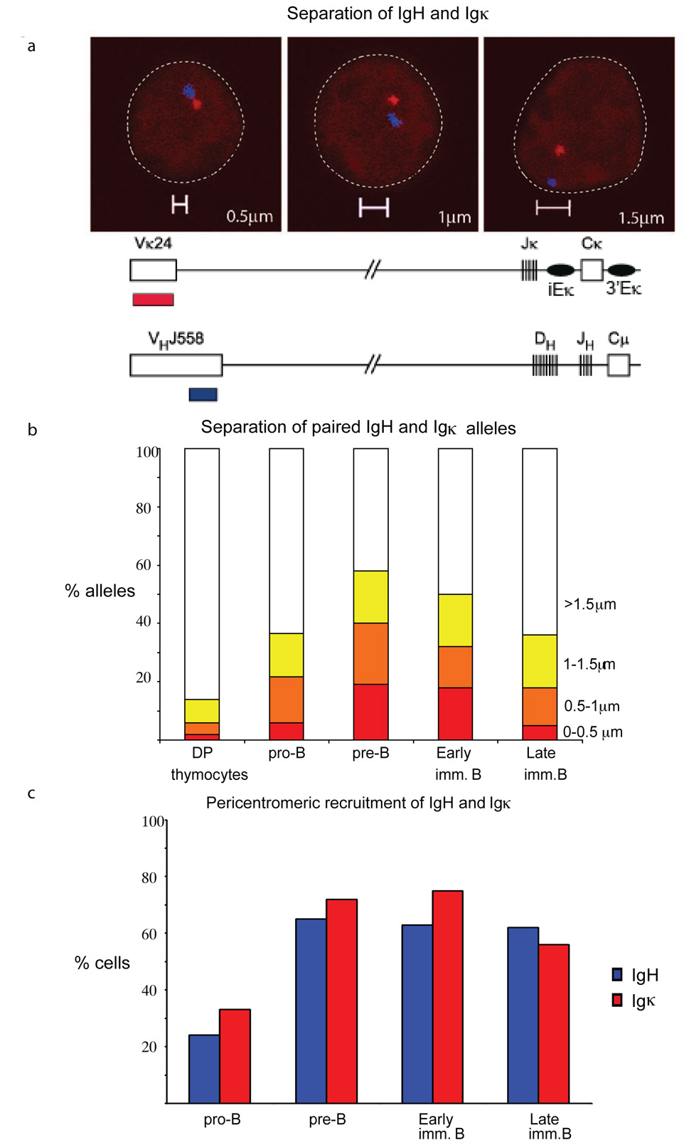
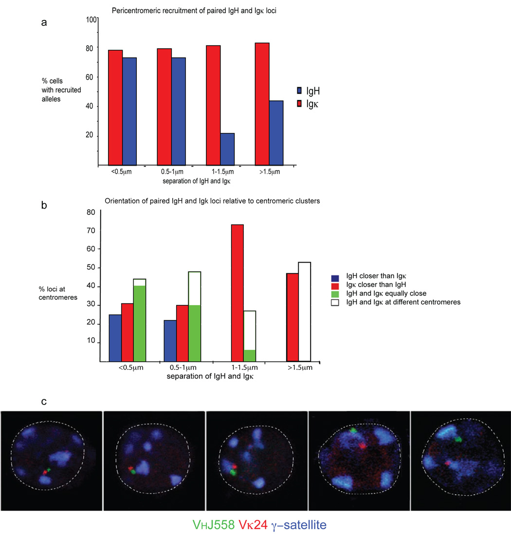
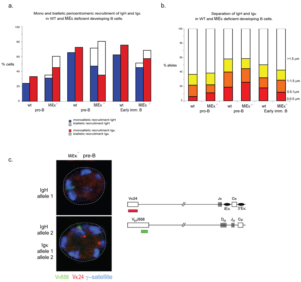
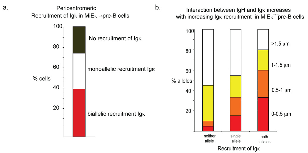
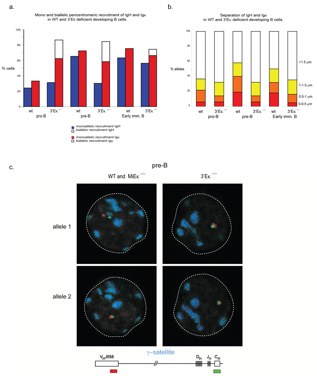
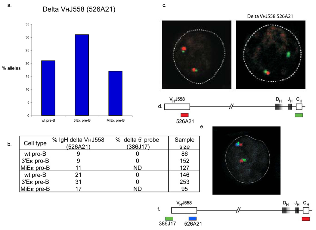
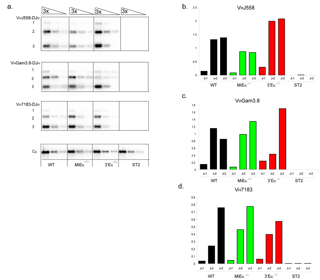
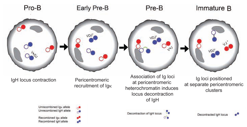
Variable-(diversity)-joining (V(D)J) recombination at loci encoding the immunoglobulin heavy chain (Igh) and immunoglobulin light chain (Igk) takes place sequentially during successive stages in B cell development. Using three-dimensional DNA fluorescence in situ hybridization, here we identify a lineage-specific and stage-specific interchromosomal association between these two loci that marks the transition between Igh and Igk recombination. Colocalization occurred between pericentromerically located alleles in pre-B cells and was mediated by the 3' Igk enhancer. Deletion of this regulatory element prevented association of the Igh and Igk loci, inhibited pericentromeric recruitment and locus 'decontraction' of an Igh allele, and resulted in greater distal rearrangement of the gene encoding the variable heavy-chain region. Our data indicate involvement of the Igk locus and its 3' enhancer in directing the Igh locus to a repressive nuclear subcompartment and inducing the Igh locus to decontract.


Figures

Figure 1
Figure 1
Interchromosomal association between one Igh and one Igk allele coincides with pericentromeric recruitment of the two loci. (a) Top, confocal sections showing distances (0.5µm, 1µm and 1.5µm) separating the 5’ ends of the Igh and Igk alleles. Bottom, schematic representation of the position of probes used for detecting Igh and Igk. (b) Distance (in µm) separating the 5’ ends of the Igh and Igk loci was evaluated in indicated subsets of sorted bone marrow cells and thymocytes. The percentage and sample sizes are shown in Supplementary Table 1, online. Cells in which no close association between Igh and Igk alleles was observed (>1.5µm) were scored as separation of a single pair of alleles. (c) Pericentromeric location of Igh and Igk alleles during B cell development. Cells at the indicated developmental stages were analyzed by three-colour3-D DNA FISH with the Igk probes RP23-101G13 (Vκ24) and RP24-387E13 (Igk constant region) in combination with a γ-satellite probe and separately with the Igh probes CT7-526A21 (VHJ558) and CT7-34H6 (CH) in combination with a γ-satellite probe. The data represent the percentage of cells showing monoallelic association of Igh (blue bars) and Igk (red bars) with pericentromeric clusters. The percentages and sample sizes of this data are shown in Supplementary Table 2, online. Data are representative of three experiments.
Figure 2
Figure 2
Association between Igh and Igk occurs on the pericentromerically recruited allele. (a) Pre-B cells were analyzed by three-color 3-dimensional DNA FISH with the Igk probe described in Fig. 1. The data represent the localization Igh and Igk alleles at different distances apart (<0.5µm, 0.5–1µm, 1–1.5µm and >1.5µm) relative to pericentromeric heterochromatin. The percentage and sample sizes are shown in Supplementary Table 3, online. (b) Evaluation of the orientation of Igh and Igk alleles at different distances apart (.0.5, 0.5–1, 1–1.5 and <1.5µm) relative to pericentromeric heterochromatin. The percentage and sample sizes are shown in Supplementary Table 4, online. (c) Confocal sections demonstrating different facets of interacting loci relative to pericentromeric heterochromatin. Data are representative of three experiments.
Figure 3
Figure 3
Increased pericentromeric recruitment of Igk promotes an increased frequency of interchromosomal association between Igh and Igkκ and increased pericentromeric recruitment of Igh dependent on 3’Eκ. (a) Pericentromeric location of Igh and Igk alleles. Cells sorted from WT and MiEκ-KO bone marrow at the indicated developmental stages were analyzed by three-color 3-dimensional DNA FISH as described for Fig. 1. The percentages and sample sizes of this data are shown in Supplementary Table 2, online.(b) The distance (in µm) separating the 5’ ends of the Igh and Igk loci was evaluated in pro-B, pre-B, IgMlow (early immature B) and IgMhigh (late immature B) cell subsets of sorted bone marrow cells from WT and MiEκ-KO mice. No IgMhigh cells were present in the mutant mice. The percentage and sample sizes are shown in Supplementary Table 1, online. (c) Top, confocal sections showing biallelically recruited Igh and Igk in pre-B cells from MiEκ-KO mice Bottom, schematic representation of the position of probes used for detecting Igh and Igk. Data are representative of three experiments.
Figure 4
Figure 4
Association of Igh and Igk increases with increasing Igk recruitment to pericentromeric regions. (a) Percentage of pre-B cells with monoallelically and biallelically pericentromeric Igk alleles, and cells where neither Igk allele is pericentromeric, in MiEκ-KO mice. The pre-B cells analyzed here were from a different batch of mice as those analyzed in the experiments shown in Fig. 3. (b) Analysis of Igh and Igk association in cells with monoallelically and biallelically pericentromeric Igk alleles, and in cells where neither Igk allele is pericentromeric. The percentage and sample sizes are shown in Supplementary Table 5, online. Data are representative of three experiments.
Figure 5
Figure 5
3’Eκ mediates association of Igh and Igk, and Igh pericentromeric recruitment at the pre-B cell stage in development. (a) Cells sorted from WT and 3’Eκ-KO bone marrow at the indicated developmental stages were analyzed by three-color 3-dimensional DNA FISH as described for Fig. 1. The percentages and sample sizes of this data are shown in Supplementary Table 2, online. (b) The distance (in µm) separating the 5’ ends of the Igh and Igk loci in pro- and pre-B cell subsets of sorted bone marrow cells from WT and 3’Eκ-KO mice. The percentage and sample sizes are shown in Supplementary Table 1, online. (c) Confocal sections showing the location of Igh alleles relative to pericentromeric clusters in pre-B cells from WT, MiEκ-KO and 3’Eκ- KO mice. The Igh probes used were CT7-526A21 (VHJ558) and CT7-34H6 (CH) in combination with a γ-satellite probe. Individual alleles from the same cell are shown in different confocal sections. Data are representative of three experiments.
Figure 6
Figure 6
Association of Igh and Igk mediates Igh locus decontraction. The distance (in µm) separating the VHJ5558 and CH gene segments in cells sorted from WT, MiEκ-KO and 3’Eκ-KO bone marrow at the indicated developmental stages were analyzed by two-color 3-dimensional DNA FISH. The Igh probes used were CT7-526A21 (VHJ558) and CT7-34H6 (CH). Data are representative of three experiments.
Figure 7
Figure 7
Deletion of the VHJ558 CT7-526A1 probe occurs more frequently in 3’Eκ-KO than in WT or MiEκ-KO pre-B cells. 3-dimensional DNA FISH was performed using the Igh probes CT7-526A21 (VHJ558) and CT7-34H6 (CH) in combination with a probe located outside of the 5’region of the Igh locus, RP24-386J17. (a) Graph showing frequency of deletion of VHJ558 526A1 in WT, 3’E-KO and MiEκ-KO pre-B cells. Table 1. showing frequency of deletion of the VHJ558 CT7-526A21 and RP24-386J17 signal in WT, 3’E κ−/− and MiE κ−/− pro-B and pre-B cells. (b) Top, confocal sections showing the presence of both CT7-526A21 (VHJ558) and CT7-34H6 (CH) signals on both Igh alleles (left) and in a different nucleus, the absence of the VHJ558 CT7-526A21 signal on one allele (right). Bottom, position and colors of the probes used. (e). Top, confocal section showing the presence of CT7-526A21 (VHJ558), CT7-34H6 (CH) and RP24-386J17 signals on one Igh allele and the absence of the VHJ558 CT7-526A21 signal on the other allele. Bottom, position and colors of the probes used. Data are representative of three experiments.
Figure 8
Figure 8
An altered spectrum of proximal versus distal VH gene segment rearrangements in 3’Eκ-KO splenic B cells. (a) PCR and Southern blot analysis of Igh rearrangements in CD19+ splenic B cells. Genotypes are indicated beneath each set of serial three-fold-diluted DNA samples. PCR primers were chosen to detect VH558 (top), VHGam3.8 (middle) and VH7183 (bottom) family rearrangements to DJH 1, 2, and 3 segments. Input DNA was normalized by amplification of a PCR fragment from the Igh Cµ region and DNA of stromal ST2 cells was used as a negative control. The numbers to the left of bands indicate rearrangements involving JH1, JH2 and JH3 gene segments. (b–d) Quantification of rearrangements shown in (a). The recombination signal is shown as a proportion of Cµ amounts for each DNA dilution and as an average across the two highest dilutions of DNA.
Figure 9
Figure 9

References

    1. Alt FW, et al. Ordered rearrangement of immunoglobulin heavy chain variable region segments. Embo J. 1984;3:1209–1219. - PMC - PubMed
    1. Jung D, Giallourakis C, Mostoslavsky R, Alt FW. Mechanism and control of V(D)J recombination at the immunoglobulin heavy chain locus. Annu Rev Immunol. 2006;24:541–570. - PubMed
    1. Kosak ST, et al. Subnuclear compartmentalization of immunoglobulin loci during lymphocyte development. Science. 2002;296:158–162. - PubMed
    1. Roldan E, et al. Locus 'decontraction' and centromeric recruitment contribute to allelic exclusion of the immunoglobulin heavy-chain gene. Nat Immunol. 2005;6:31–41. - PMC - PubMed
    1. Geier JK, Schlissel MS. Pre-BCR signals and the control of Ig gene rearrangements. Semin Immunol. 2006;18:31–39. - PubMed

Publication types

MeSH terms

LinkOut - more resources