. 2008 Feb 20;3(2):e1647.
doi: 10.1371/journal.pone.0001647.

Golgi cisternal unstacking stimulates COPI vesicle budding and protein transport

Affiliations

Golgi cisternal unstacking stimulates COPI vesicle budding and protein transport

Yanzhuang Wang et al. PLoS One. .

Abstract

The Golgi apparatus in mammalian cells is composed of flattened cisternae that are densely packed to form stacks. We have used the Golgi stacking protein GRASP65 as a tool to modify the stacking state of Golgi cisternae. We established an assay to measure protein transport to the cell surface in post-mitotic cells in which the Golgi was unstacked. Cells with an unstacked Golgi showed a higher transport rate compared to cells with stacked Golgi membranes. Vesicle budding from unstacked cisternae in vitro was significantly increased compared to stacked membranes. These results suggest that Golgi cisternal stacking can directly regulate vesicle formation and thus the rate of protein transport through the Golgi. The results further suggest that at the onset of mitosis, unstacking of cisternae allows extensive and rapid vesiculation of the Golgi in preparation for its subsequent partitioning.


Conflict of interest statement

Competing Interests: The authors have declared that no competing interests exist.

Figures

Figure 1
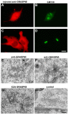
Figure 1. Microinjection of antibodies to GRASP65 or recombinant G2A GRASP65 prevents post-mitotic Golgi stack formation.
(A–D) NRK cells in metaphase were microinjected with affinity-purified antibodies to GRASP65. The cells were fixed either immediately (A, B) or after cell division (C, D). The injected antibodies (A, C) and the Golgi marker GM130 (B, D) were stained. The microinjected cells divided into two daughter cells, each of which has a reformed Golgi ribbon. Bar, 15 µm. (E–H) NRK cells in metaphase were microinjected with anti-GRASP65 (E, F), G2A GRASP65 protein (G), or non-specific rabbit IgGs as a control (H). FITC-labeled dextran was co-injected as a marker. Once cell division was complete, all non-injected cells were removed and the remaining injected cells were processed for EM. Shown are EM micrographs of the reassembled Golgi in the injected cells. Note that in control IgG injected cells, the Golgi stacks are properly reassembled. In cells injected with either anti-GRASP65 or G2A GRASP65, however, cisternae appear disorganized and poorly aligned. In close proximity to the cisternae, there are a large number of vesicles and budding profiles, which appear to be coated. Bar, 0.5 µm.
Figure 2
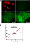
Figure 2. CD8 transport assay.
(A) A plasmid encoding the plasma membrane protein CD8 was microinjected together with Texas-Red dextran (top left panel, red) into NRK cells. After 45 min, a fluorescently labeled antibody against the luminal domain of CD8 was added to the medium. The recruitment of the antibody onto the cell surface was analyzed by time-lapse microscopy in 2 min intervals over 70 min (Supplemental Movie S1). The fluorescence intensity on the cell surface increased over time, representing the arrival of CD8 from the Golgi to the plasma membrane. Bar, 15 µm. (B) Quantitation. To validate the transport assay, a peptide (N73) that inhibits intra-Golgi transport, or a control peptide (wt), were co-injected into NRK cells along with the CD8 plasmid and Texas-Red dextran and assayed for CD8 transport as in (A). The mean fluorescence intensity of the cells in each frame was quantified and plotted (red curve: control, n = 10; black curve: N73 peptide, n = 6); it decreased from 9.7 per min for control-injected cells to 2.4 per min for N73-peptide injected cells. CD8 transport was therefore inhibited by 75%.
Figure 3
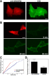
Figure 3. CD8 transport to the plasma membrane is accelerated through unstacked Golgi membranes.
(A) Metaphase NRK cells were microinjected with affinity-purified antibodies against GRASP65 together with a plasmid encoding the plasma membrane protein CD8. After cell division, CD8 was expressed in the daughter cells and transported to the plasma membrane. 45 min before fixation, cycloheximide was added to inhibit protein translation. Cells were double labeled with a monoclonal antibody against CD8 and the microinjected polyclonal antibodies against GRASP65. Note that CD8 was transported to the cell surface in cells where Golgi stack formation was inhibited by the injected GRASP65 antibodies. Bar, 15 µm. (B) Metaphase NRK cells were injected with G2A GRASP65 protein together with a CD8 plasmid and Texas-Red dextran as a marker. At the end of mitosis, a fluorescently labeled antibody against CD8 was added to the medium. The fluorescence increase was followed by time-lapse microscopy in 2 min intervals over 70 min (Movie S2). Bar, 15 µm. (C) Quantitation. The mean fluorescence intensity of the cells in each frame was quantified and plotted (red curve: G2A GRASP65 injection, blue curve: control injection). The data shown were obtained from one pair of divided cells for each condition. Note the increased rate of transport in cells injected with G2A GRASP65. (D) The transport rate of CD8 to the plasma membrane increased two-fold in cells with unstacked Golgi cisternae. Quantitation from three independent experiments (mean±SD; p<0.05). Appearance of CD8 per minute at the cell surface was stimulated by 90% in G2A GRASP65 injected cells (n = 34) compared to control cells (n = 44).
Figure 4
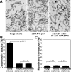
Figure 4. Inhibition of stacking by antibodies against GRASP65.
(A) Rat liver Golgi membranes (RLG) were incubated with cdk1/cyclin B1 and plk1 and fixed either directly or after further incubation with interphase HeLa cell cytosol in the presence of GRASP65 antibodies. Representative EM micrographs of the membranes are shown in (A). Incubation with mitotic kinases led to unstacking of Golgi cisternae (cdk1/cyclin B1+plk1), and the subsequent reformation of stacks upon treatment with interphase cytosol was inhibited by antibodies against GRASP65. Bar, 0.5 µm. (B, C) Quantitation of (A) by the intersection method (mean±SD). Upon treatment with mitotic kinases, membranes in stacks were reduced to 11%, compared to 71% for untreated Golgi membranes. When interphase single cisternae were generated in the presence of GRASP65 antibodies and interphase cytosol, the percentage of membranes in stacks remained unchanged (B). During this process, the single cisternal membranes remained intact and did not vesiculate, as the percentage of membranes in vesicles was unchanged between all groups (C). Statistical significance was assessed by a two-tailed Student's t test.
Figure 5
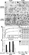
Figure 5. Unstacking of Golgi cisternal membranes increases vesicle generation.
(A–D) EM micrographs of untreated Golgi membranes (A), or interphase single cisternae (C) generated by sequential treatment with cdk1/cyclin B1 and plk1 and interphase cytosol in the presence of GRASP65 antibodies. Membranes in (A) and (C) were treated with coatomer and ARF1 to allow vesicles to form. Membranes were fixed at different time points and processed for EM. Shown in (B) and (D) are membrane profiles after 5 min incubation. Note that budding from single cisternae was increased compared to that from stacked membranes. Within 5 min, nearly all single cisternae were converted into vesicles (D), while budding from stacked cisternal membranes was less efficient (B). Bar, 0.5 µm. (E) Quantitation of (A–D). Untreated Golgi membranes as in A, or single cisternae as in C were incubated with coatomer and ARF1 and then analyzed by EM. Shown is the quantitation of EM micrographs by the intersection method. The rate of vesicle formation from single cisternae was doubled (t1/2 = 1.6 min) compared to stacked membranes (t1/2 = 3.5 min). 78.5% of the membranes from single cisternae were converted into vesicles with a half-time of 1.6 min. The formation of the same amount of vesicles from stacked membranes took 12.4 min. Statistical significance assessed by a two-tailed Student's t test showed that p<0.01 for all time points except 0 min. (F) Untreated Golgi membranes (c, Golgi stacks) were treated with cdk1/cyclin B1 and plk1 followed by further incubation with interphase cytosol in the presence (a, unstacked cisternae) or absence (b, restacked cisternae) of GRASP65 antibodies. These membranes were incubated with coatomer and ARF1 for the indicated times followed by separation of the Golgi membranes (G) and vesicles (V) by centrifugation. Equal fractions of the samples were analyzed by Western blotting for ManII and Gos28. (G) Quantitation of (F). ManII was recruited more efficiently into the vesicle fractions from single cisternae compared to stacks or restacked membranes, whereas the t-SNARE Gos28 remained in the Golgi fraction. Results represent 3 independent experiments.

Similar articles

Cited by

References

    1. Marsh BJ, Volkmann N, McIntosh JR, Howell KE. Direct continuities between cisternae at different levels of the Golgi complex in glucose-stimulated mouse islet beta cells. Proc Natl Acad Sci U S A. 2004;101:5565–5570. - PMC - PubMed
    1. Mironov AA, Beznoussenko GV, Polishchuk RS, Trucco A. Intra-Golgi transport: a way to a new paradigm? Biochim Biophys Acta. 2005;1744:340–350. - PubMed
    1. Trucco A, Polishchuk RS, Martella O, Di Pentima A, Fusella A, et al. Secretory traffic triggers the formation of tubular continuities across Golgi sub-compartments. Nat Cell Biol. 2004;6:1071–1081. - PubMed
    1. Elsner M, Hashimoto H, Nilsson T. Cisternal maturation and vesicle transport: join the band wagon! (Review). Mol Membr Biol. 2003;20:221–229. - PubMed
    1. Losev E, Reinke CA, Jellen J, Strongin DE, Bevis BJ, et al. Golgi maturation visualized in living yeast. Nature. 2006;441:1002–1006. - PubMed

Publication types