. 2008 Sep;57(9):1381-90.
doi: 10.1007/s00262-008-0474-9. Epub 2008 Feb 23.

CD54 is a surrogate marker of antigen presenting cell activation

Affiliations

CD54 is a surrogate marker of antigen presenting cell activation

N A Sheikh et al. Cancer Immunol Immunother. 2008 Sep.

Abstract

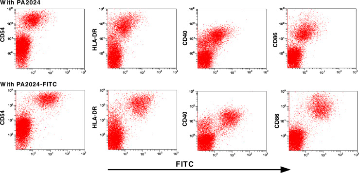
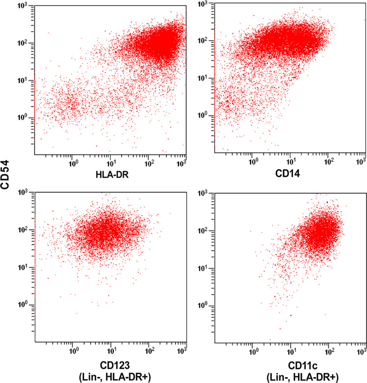
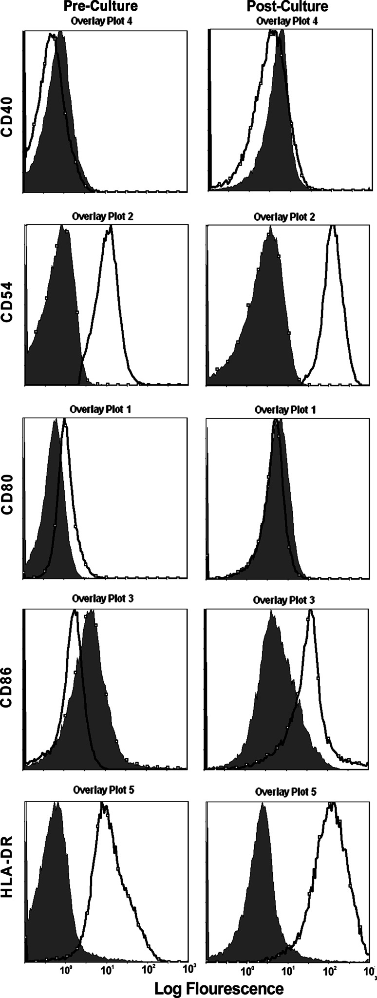
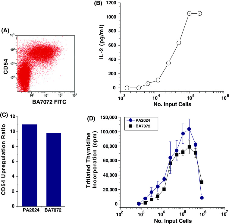
There is no single universally accepted hallmark of antigen presenting cell (APC) activation. Instead a variety of methods are used to identify APCs and assess their activation state. These activation measures include phenotypic methods [e.g., assessing the increased expression of surface markers such as major histocompatability (MHC) class II] and functional assays (e.g., evaluating the enhanced ability to take up and process antigen, or stimulate naïve T cells). Sipuleucel-T is an investigational autologous active cellular immunotherapy product designed to stimulate a T cell immune response against human prostatic acid phosphatase (PAP), an antigen highly expressed in prostate tissue. Sipuleucel-T consists of peripheral blood mononuclear cells (PBMCs), including activated APCs displaying epitopes of PAP. In order to develop a robust reproducible potency assay that is not hampered by MHC restriction we have developed a method to simply assess the biological activation of antigen presenting cells (APCs). In the course of sipuleucel-T characterization, we analyzed various phenotypic and functional parameters to define the activation state of APCs obtained from peripheral blood. Flow cytometric assays revealed that CD54+ cells are responsible for antigen uptake, and that expression of CD54 predominantly localizes to APCs. Costimulation, as measured by an allogeneic mixed lymphocytic reaction (alloMLR) assay, showed that activity was restricted to the CD54+ cell population. Similarly, CD54+ cells harbor all of the PAP-specific antigen presentation activity, as assayed using a PAP-specific HLA-DRbeta1-restricted T cell hybridoma. Finally we show that CD54 expression is substantially and consistently upregulated on APCs during culture with a GM-CSF fusion protein, and that this upregulation activity can be quantified. Thus these data support the use of CD54 upregulation as a surrogate for assessing human APC activation and validates its utility as a potency measure of sipuleucel-T.


Figures

Fig. 1
Fig. 1
FITC labeled PA2024 is assimilated preferentially by CD54+ cells. Cells were incubated with ether PA2024 or PA2024 spiked with 1% (w/w) FITC labeled PA2024 and after approximately 40 h cells were harvested as described in the materials and methods and surface stained for CD54, HLA-DR, CD40 and CD86. Stained cells were analyzed on a BD FACSAria™ flow cytometer and data is displayed using CXP software. The top row of flow plots represents cells cultured with unlabeled PA2024 and the lower row is from cells cultured with PA2024 spiked with FITC labeled PA2024. Data presented is from one representative set from multiple experiments
Fig. 2
Fig. 2
CD54+ cells assimilate and present antigen. a Cells were incubated with PA2024 spiked with 1% (w/w) FITC labeled PA2024. After approximately 40 h cells were harvested and sorted on the basis of their FITC signal as described in Materials and methods. The two resultant cell populations (FITC+ and FITC−) were evaluated for expression of PAP epitopes using the PAP-specific Papillon hybridoma. FITC+ and FITC− cells were titrated in triplicate against 1 × 105 Papillon cells in 96 well flat bottomed plates. IL-2 production was measured by ELISA. b Cells cultured with PA2024 were sorted on the basis of CD54 expression. CD54+and CD54- cells were titrated in triplicate against 1 × 105 Papillon cells in 96 well flat bottomed plates and IL-2 production was measured by direct ELISA. Data presented is from one representative set from multiple experiments
Fig. 3
Fig. 3
Antigen presenting cells express CD54. Post culture cells were co-stained with anti-CD54 PE, anti-CD14 PerCPCy5.5, anti-HLA-DR APC-Cy7 or with anti-lineage cocktail-FITC in combination with anti-CD54-PE, anti-HLA-DR APC-Cy7, anti-CD11c APC and anti-CD123 PerCP Cy5.5. Stained cells were collected on a BD FACSAria™ flow cytometer where a total of 500,000 events were collected. Analysis was restricted to large cells (determined by forward scatter vs side scatter). In the case of cells stained for myeloid (CD11c) and plasmacytoid (CD123) DC markers analysis was based upon large cells that were lineage negative and HLA-DR positive. Data presented is a representative set from multiple experiments
Fig. 4
Fig. 4
CD54+ cells possess costimulatory activity, which increases after culture with the fusion protein PA2024. Prior to and immediately after culture with antigen, cells were sterile sorted on a BD FACSAria on the basis of CD54 expression. CD54+ sorted cells were irradiated and titrated in 96 well flat bottom plates in quadruplicate against 5 × 104 human CD3+ cells purified from multiple healthy donors. Plates were incubated for 6 days at 37°C with a pulse of 0.5 mCi [3H] thymidine for the final 18 h. Cells were harvested and read on a betaplate counter. CD54- sorted cells did not have any stimulatory capacity as evidenced by the fact that incorporation of tritiated thymidine was below background levels (not presented). The data displayed is from one of two independent representative experiments
Fig. 5
Fig. 5
CD54 expression is increased after culture with PA2024 antigen. Cells were incubated with PA2024 for approximately 40 h. Cells were stained with anti-CD80 FITC, CD54 PE, CD86 PE-Cy5, CD40 APC, HLA-DR APC-Cy7 and run on a BD FACSAria where 200,000 events were collected. The shaded peaks represent staining obtained by isotype-matched fluorescent-tagged antibodies and the thick black lines are staining with the surface marker indicated on the Y axis for each pair of histograms. Data displayed is from one of seven experiments
Fig. 6
Fig. 6
CD54+ cells take up BA7072 and surface CD54 is upregulated. a Cells were incubated with BA7072 spiked with 1% (w/w) FITC labeled BA7072. After approximately 40 h cells were harvested as described in the materials and methods and surface stained for CD54. Stained cells were analyzed on a BD FACSAria™ flow cytometer and data is displayed using CXP software. b Cells cultured with BA7072 process and present Her2/neu epitopes to Herder, a Her2/neu-specific T cell hybridoma. As described in Materials and methods, post culture cells were titrated against Herder hybridoma and the level of Herder activation was assessed by measuring the amount of IL-2 produced. c CD54 is upregulated after culture with either BA7072 or PA024. Surface CD54 was measured on pre and post culture cells and the relative increase in CD54 expression was reported as a ratio. d Cells cultured with either BA7072 or PA2024 possess costimulatory activity. Post culture cells were irradiated and incubated with pooled purified human T cells as described previously and the degree of T cell proliferation was inferred from the amount of tritiated thymidine incorporation

References

    1. Adams DO, Hamilton TA. The cell biology of macrophage activation. Annu Rev Immunol. 1984;2:283–318. doi: 10.1146/annurev.iy.02.040184.001435. - DOI - PubMed
    1. Burch PA, Breen JK, Buckner JC, Gastineau DA, Kaur JA, Laus RL, Padley DJ, Peshwa MV, Pitot HC, Richardson RL, Smits BJ, Sopapan P, Strang G, Valone FH, Vuk-Pavlovic S. Priming tissue-specific cellular immunity in a phase I trial of autologous dendritic cells for prostate cancer. Clin Cancer Res. 2000;6:2175–2182. - PubMed
    1. Burch PA, Groghan GA, Gastineau DA, Jones LA, Kaur JS, Kylstra JW, Richardson RL, Valone FH, Vuk-Pavlovic S. Immunotherapy (APC8015, Provenge) targeting prostatic acid phosphatase can induce durable remission of metastatic androgen-induced prostate cancer: a phase 2 trial. Prostate. 2004;60:197–204. doi: 10.1002/pros.20040. - DOI - PubMed
    1. Carrasco YR, Fleire SJ, Cameron T, Dustin ML, Batista FD. LFA-1/ICAM-1 interaction lowers the threshold of B cell activation by facilitating B cell adhesion and synapse formation. Immunity. 2004;20:58–599. doi: 10.1016/S1074-7613(04)00105-0. - DOI - PubMed
    1. Coyle AJ, Gutierrez-Ramos JC. The expanding B7 superfamily: increasing complexity in costimulatory signals regulating T cell function. Nat Immunol. 2001;2:203–209. doi: 10.1038/85251. - DOI - PubMed
Online references
    1. From a presentation at the Cellular, Tissue and Gene Therapies Advisory Committee meeting. January 2007 posting date. Gavin D. Perspectives on potency assays for complex biological products. Food and Drug Administration, Center for Biologics Research and Evaluation, Office of Cellular, Tissue and gene Therapy. http://www.fda.gov/cber/genetherapy/cmc012807dg.htm

MeSH terms