. 2008 May;29(5):905-12.
doi: 10.1093/carcin/bgn049. Epub 2008 Feb 24.

Expression of the type III TGF-beta receptor is negatively regulated by TGF-beta

Affiliations

Expression of the type III TGF-beta receptor is negatively regulated by TGF-beta

Nadine Hempel et al. Carcinogenesis. 2008 May.

Abstract

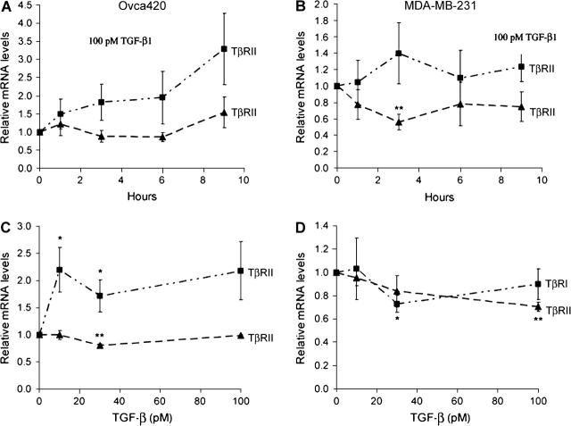
The type III transforming growth factor-beta receptor (TbetaRIII or betaglycan) is a ubiquitously expressed transforming growth factor-beta (TGF-beta) superfamily coreceptor with essential roles in embryonic development. Recent studies have defined a role for TbetaRIII in the pathogenesis of human cancers, with frequent loss of TbetaRIII expression at the message and protein level. Mechanisms for the loss of TbetaRIII expression remain to be fully defined. Advanced human cancers often have elevated circulating levels of TGF-beta1. Here, we define a specific role for TGF-beta1 in negatively regulating TbetaRIII at the message level in breast and ovarian cancer models. TGF-beta1 decreased TbetaRIII message and protein levels in ovarian (Ovca420) and breast cancer (MDA-MB-231) cell lines in both a dose- and time-dependent manner. TGF-beta1-mediated TbetaRIII repression is mediated by the type I TGF-beta receptor/Smad2/3 pathway as the activin receptor-like kinase 5 (ALK5) inhibitor, SB431542, abrogated this effect, while the expression of constitutively active ALK5 was sufficient to repress TbetaRIII expression. Mechanistically, TGF-beta1 does not affect TbetaRIII messenger RNA (mRNA) stability, but instead directly regulates the TbetaRIII promoter. We define alternative promoters for the TGFBR3 gene, a distal and proximal promoter. Although both promoters are active, only the proximal promoter was responsive and negatively regulated by TGF-beta1 and constitutively active ALK5. Taken together, these studies define TGF-beta1-mediated downregulation of TbetaRIII mRNA expression through effects on the ALK5/Smad2/3 pathway on the TGFBR3 gene proximal promoter as a potential mechanism for decreased TbetaRIII expression in human cancers.


Figures

Fig. 1.
Fig. 1.
TGF-β1 downregulates TβRIII expression. Ovca420 (A) and MDA-MB-231 (B) cells were treated 100 pM TGF-β1 for the indicated times. For dose-response experiments Ovca420 (C) and MDA-MB-231 (D) cells were treated for 6 hours with the indicated amounts of TGF-β1. RNA was isolated, reverse transcribed and Real time semi-quantitative PCR carried out with primers against human TβRIII. Data was normalized against GAPDH levels and expressed relative to fold reduction in TβRIII levels compared to non-treated controls (n = 3, mean +/− SEM, *P < 0.05, **P < 0.01, ***P < 0.001, student's T-test). E. Ovca420 and MDA-MB-231 cells were treated for 24 hours with the indicated amounts of TGF-β1 (non-treated controls: NT), and TβRIII protein levels were detected by binding and crosslinking of 125I-TGF-β1 to the receptor. TβRIII was immunoprecipitated from lysed cells (cell surface) or media (soluble) using TβRIII specific antibodies, followed by protein electrophoresis and visualization using autoradiography. The arrow points to the position of TβRIII on the gel and the position of the 165 kDa molecular weight marker is indicated.
Fig. 2.
Fig. 2.
TGF-β1 does not significantly regulate TβRI and TβRII expression. Ovca420 (A) and MDA-MB-231 (B) cells were treated 100 pM TGF-β1 for indicated times. For dose-response experiments Ovca420 (C) and MDA-MB-231 (D) cells were treated for 6 hours with indicated amounts of TGF-β1. RNA was isolated, reverse transcribed and Real time semi-quantitative PCR carried out with primers against human TβRI and TβRII. Data was normalized against GAPDH levels and expressed relative to fold changes in TβRI or TβRII levels compared to non-treated controls (n = 3, mean +/− SEM, *P < 0.05, **P < 0.01, student's T-test).
Fig. 3.
Fig. 3.
TGF-β acts via TβRI (ALK5) to down-regulate TβRIII expression. A. Ovca420 and MDA-MB-231 cells were pretreated with the ALK5 inhibitor SB431542 (20 μM) for 30 min, following by 6 hours of 100 pM TGF-β1 treatment. RNA was isolated, reverse transcribed and real time semi-quantitative RT-PCR carried out with TbRIII specific primers. Values were normalized against GAPDH levels and expressed relative to fold reduction in TβRIII levels (n = 3, mean +/− SEM, **P < 0.01, ***P < 0.001, student's T-test). B. MDA-MB-231 cells were infected with adenoviral construct expressing the constitutively active form of ALK5 (ALK5QD) or control adenovirus expressing GFP. 48 hours post infection RNA was isolated, reverse transcribed and real time semi-quantitative RT-PCR carried out with TβRIII specific primers. Values were normalized against GAPDH levels and expressed relative to fold reduction in TβRIII levels (n = 2, mean +/− SEM, *P < 0.05, student's T-test).
Fig. 4.
Fig. 4.
TGF-β1 does not alter TβRIII mRNA stability. Ovca420 (A) and MDA-MB-231 (B) cells were treated with 10 μg/ml Actinomycin D and with or without 100pM TGF-β1 for the indicated times. RNA was isolated, reverse transcribed and real time semi-quantitative RT-PCR carried out with TβRIII specific primers. Values were normalized against GAPDH levels and expressed relative to fold reduction in TβRIII levels (n = 3, mean +/− SEM).
Fig. 5.
Fig. 5.
Activity of the proximal and distal promoters of the TGFBR3 gene. A. Schematic of the 5′ region of the human TGFBR3 gene. Grey boxes represent the location of the alternate 5′UTRs of TβRIII cDNA species identified in the literature and black box the first coding exon of TβRIII. The 5′UTR transcriptional start sites of the proximal and distal promoters are labeled as +1. The location of the promoter constructs and potential transcription factor binding sites are indicated B. Promoter activities of sequences flanking the alternate 5′UTRs of TβRIII in Ovca420 and MDA-MB-231 cells. All promoters were cloned into the pGL3Basic vector, transfected into cells and lysed cells assayed for luciferase activity as stated in Methods. Results are corrected for Renilla luciferase activity of the pRLSV40 transfection standard and represent the mean +/− SEM, n = 3. Results are expressed as fold increases in luciferase activity relative to the empty pGL3Basic vector.
Fig. 6.
Fig. 6.
The proximal TβRIII promoter is negatively regulated by TGF-β1. A. The empty pGL3Basic vector, the TGF-β responsive p3TP promoter, the proximal (−559/+60) and the distal (−2330/+80) TβRIII promoter constructs were transfected into Ovca420 cells and treated for 48 hours with 100pM TGF-β1. Cells were lysed and assayed for luciferase activity as stated in Methods. Results are corrected for Renilla luciferase activity of the pRLSV40 transfection standard and represent the mean +/− SEM, n = 3. Results are expressed as fold changes in luciferase activity relative to each untreated control (NT, **P<0.01, ***P<0.001, student's T-test). B. TGF-β down-regulates TβRIII proximal promoter activity in a dose-dependent manner. Ovca420 cells were transfected with proximal and distal promoter as stated above and treated with indicated amounts of TGF-β1 for 48 hours. Results are expressed as fold changes in luciferase activity relative to each untreated control (0 pM; mean +/− SEM, n = 3, *P<0.05, **P<0.01, student's T-test). C. TβRIII proximal promoter downregulation is ALK5-dependent. Constitutive active ALK5QD was cotransfected with either the p3TP promoter construct, the TβRIII proximal or distal promoter into Ovca420 cells. 48 hours post-transfection luciferase activity was measured as stated above and treated with indicated amounts of TGF-β1 for 48 hours. Results are expressed as fold changes in luciferase activity relative to each untransfected control (NT; mean +/− SEM, n = 3, **P<0.01, student's T-test).

References

    1. Elliott RL, et al. Role of transforming growth factor Beta in human cancer. J. Clin. Oncol. 2005;23:2078–2093. - PubMed
    1. Wrana JL, et al. Mechanism of activation of the TGF-beta receptor. Nature. 1994;370:341–347. - PubMed
    1. Nakao A, et al. TGF-beta receptor-mediated signalling through Smad2, Smad3 and Smad4. EMBO J. 1997;16:5353–5362. - PMC - PubMed
    1. Lopez-Casillas F, et al. Betaglycan presents ligand to the TGF beta signaling receptor. Cell. 1993;73:1435–1444. - PubMed
    1. Lewis KA, et al. Betaglycan binds inhibin and can mediate functional antagonism of activin signalling. Nature. 2000;404:411–414. - PubMed

Publication types

MeSH terms