. 2008 Apr;36(7):2338-52.
doi: 10.1093/nar/gkn068. Epub 2008 Feb 24.

Requirements for gene silencing mediated by U1 snRNA binding to a target sequence

Affiliations

Requirements for gene silencing mediated by U1 snRNA binding to a target sequence

Xabi Abad et al. Nucleic Acids Res. 2008 Apr.

Abstract

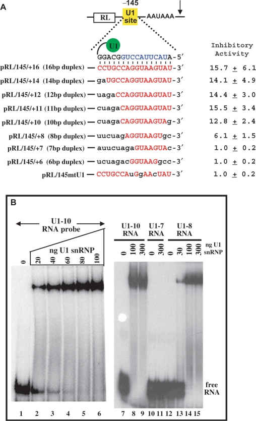
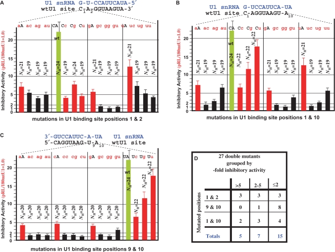
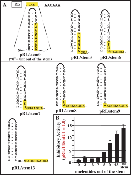
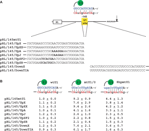
U1 interference (U1i) is a novel method to block gene expression. U1i requires expression of a 5'-end-mutated U1 snRNA designed to base pair to the 3'-terminal exon of the target gene's pre-mRNA that leads to inhibition of polyadenylation. Here, we show U1i is robust (> or =95%) and a 10-nt target length is sufficient for good silencing. Surprisingly, longer U1 snRNAs, which could increase annealing to the target, fail to improve silencing. Extensive mutagenesis of the 10-bp U1 snRNA:target duplex shows that any single mismatch different from GU at positions 3-8, destroys silencing. However, mismatches within the other positions give partial silencing, suggesting that off-target inhibition could occur. The specificity of U1i may be enhanced, however, by the fact that silencing is impaired by RNA secondary structure or by splicing factors binding nearby, the latter mediated by Arginine-Serine (RS) domains. U1i inhibition can be reconstituted in vivo by tethering of RS domains of U1-70K and U2AF65. These results help to: (i) define good target sites for U1i; (ii) identify and understand natural cellular examples of U1i; (iii) clarify the contribution of hydrogen bonding to U1i and to U1 snRNP binding to 5' splice sites and (iv) understand the mechanism of U1i.


Figures

Figure 1.
Figure 1.
Inhibitory activity of U1 snRNAs having extended 5′ends. (A). Exogenous 5′-end mutated U1 snRNA inhibits the expression of the targeted Renilla reporter in a dose-dependent manner. HeLa cells were cotransfected with a Renilla pRL/87mtU1 reporter that contains a mtU1 binding site at position –87 and variable amounts (1, 2 or 3 µg) of mtU1/+0 plasmid that expresses a 5′-end mutated U1 snRNA designed to base pair to the mtU1-binding site as indicated. ‘+0’ serves to indicate a normal length 5′ end as opposed to an extended 5′ end. As a control, a plasmid expressing wtU1 was used in place of the mtU1/+0 plasmid. In all cases, a firefly plasmid was also cotransfected for normalization purposes. The inhibitory activity of the mtU1/+0 plasmid was calculated by dividing the normalized Renilla activity of the transfection with the pRL/87mtU1 + wtU1 plasmids by the normalized Renilla activity of the transfection with the pRL/87mtU1 + mtU1/+0 plasmids. The wtU1 plasmid did not affect expression of pRL/87mtU1 and therefore had an inhibitory value set to 1.0. The standard deviations were calculated from three independent experiments. (B) 5′ end extended U1 snRNAs do not increase inhibitory activity. The plasmid that expresses mtU1/+0 was used as a parental vector to construct plasmids that express U1 snRNA extended at the 5′-end +1, +2, +3, +4, +5, +6 and +15 nt (mutU1/+1, mutU1/+2, etc.). These extensions increase the length of the duplex formed between pRL/87mtU1 pre-mRNA and the mtU1-derived snRNA. The sequence of the 5′ end of the U1 snRNA and the duplex length is indicated for each case. As described in (A), all plasmids were transfected along with pRL/87mtU1 and firefly luciferase and their relative inhibitory activities were calculated. The inhibitory activities are derived from five different experiments. Standard deviations are not shown but were <25% in each case. (C) 5′-end extended snRNAs are stably expressed. A primer extension reaction was performed with no RNA (lane 1) or with total RNA isolated from cells transfected with the wtU1 expressing plasmid (lane 2) or the mtU1-derived plasmids from (B) as indicated (lane 3–10). The 32P-end-labeled oligo used recognizes both endogenous wtU1 and all mtU1-derived snRNAs, as indicated to the right of the figure. The samples were separated by denaturing PAGE and visualized by autoradiography. The product obtained with mtU1/+0 snRNA in lane 3 comigrates with the product of endogenous U1 snRNA. The product of mtU1/+1 snRNA in lane 4 is difficult to visualize because it would be only 1-nt longer than the product of endogenous U1 snRNA.
Figure 2.
Figure 2.
Effect of U1-binding site:U1 snRNA duplex length on inhibition. (A) Shown is a series of Renilla luciferase plasmids with different U1-binding sites cloned at 145 nt from the poly(A) signal (pRL/145/x). Shown in red are nucleotides from the U1-binding site able to bind endogenous U1 snRNA. The U1-binding site:U1 snRNA duplex length is indicated for each case. As was done in the Supplementary Data Figure S1, HeLa cells were transfected with these plasmids along with a firefly luciferase plasmid for normalization purposes. Inhibitory activities were calculated as in the Supplementary Data Figure S1 and the bar graph summarizes five independent experiments. The pRL/145mtU1 is the reference control plasmid that matches pRL/145/+16 except for three point mutations in the U1-binding site. (B) An electrophoretic mobility shift assay (EMSA) was used to detect binding of purified U1 snRNP to 0.03 pmol 32P-radiolabeled RNAs with various types of U1-binding sites. EMSA conditions were as previously described (18). The RNAs are all matching except for differences in the U1-binding site sequence. Lanes 1–9 contain 32P labeled U1-10 RNA that has a 10-nt wt U1-binding site. Lanes 10 and 11 contain U1-7 RNA that matches U1-10 RNA except the U1-binding site is 7-nt long. Lanes 12–15 contain U1-8 RNA that matches U1-10 RNA except the U1-binding site is 8-nt long. The amounts of purified U1 snRNP added are indicated (note 100 ng U1 snRNP = 0.3 pmol). The purification of U1 snRNP is described in the Supplementary Data Figure S2. The experiment was repeated ×5, quantitated by phosphorimagery and a kDa of 4 +/− 1.5 nM was calculated for the U1 snRNP:U1-10 RNA complex. The U1-8 RNA bound about 3× weaker than the U1-10 RNA and no detectable binding to the U1-7 RNA was observed under these conditions.
Figure 3.
Figure 3.
Analysis of single-point mutations of the U1-binding site. A saturation mutagenesis analysis was performed where all 30 possible single-point mutations were introduced in the 10-nt wtU1-binding site of the pRL/180wtU1 plasmid shown in the Supplementary Data Figure S1. Each plasmid was cotransfected with the firefly luciferase control into HeLa cells and inhibitory activities were calculated as described in the Supplementary Data Figure. S1. The results are plotted in a bars graph where error bars indicate standard deviations of five independent experiments. The sequence of the wtU1-binding site and its base pairing with the 11 nt of the 5′-end of U1 snRNA (blue font) is schematized above the graph. Each dotted line indicates the mutation (in red font) which is positioned above its corresponding bar graph representing the inhibitory activity of that mutation. The inhibitory activity of the reporter with a wtU1-binding site is shown in green to the left of the graph. The 6G and 7G single mutants were combined to give the 6G/7G double mutant whose activity is shown on the far right.
Figure 4.
Figure 4.
Analysis of U1-binding sites with double-point mutations at positions 1 and 2, 1 and 10 or 9 and 10. A saturation mutagenesis analysis was performed where all possible double mutations were introduced at positions 1 and 2 (A) 1 and 10 (B) and 9 and 10 (C) of the 10-nt wtU1-binding site of the pRL/180wtU1 plasmid shown in the Supplementary Data Figure S1. The mutations were analyzed and graphed as in Figure 3. The sequence of the wtU1-binding site and its base pairing with the 11 nt of the 5′-end of U1 snRNA (blue font) is schematized above the graph. Each pair of letters indicates a particular mutant where the lowercase red letters correspond to a mutation, while the uppercase black letters match the wtU1-binding site. Each pair of letters is positioned above its corresponding bar graph representing the inhibitory activity of that mutated U1 site. Also indicated are the NH values that are defined as the number of continuous hydrogen bonds (see Discussion section). To facilitate comparison we included the single-point mutations from Figure 2 as red histograms. All double mutants (totals) or double mutants from (A) (1 and 2), (B) (1 and 10) and (C) (1 and 10) were classified according to their good, low or no inhibitory activity (>5-, 2–5- and 2-fold inhibition, respectively). Shown is the number of double mutants in each group (D).
Figure 5.
Figure 5.
Effect of secondary structure on U1-binding site inhibition. (A) The pRL/145/stem0 plasmid has a 13-nt wtU1-binding site (the canonical 10 nt are highlighted in yellow) completely occluded in a stem–loop sequence. The ‘0’ indicates 0 nt of the U1-binding site should be found outside of the stem. As diagrammed, a collection of plasmids were made and tested that match pRL/145/stem0 except the U1-binding site increasingly moves out of the stem. The number below each stem–loop structure indicates the number of U1-binding site nucleotides that should be found outside of the stem (3, 6, 7, 8, 9 or 13). (B) The plasmids were analyzed and graphed as indicated in Figure 3. Error bars indicate standard deviations of four different experiments.
Figure 6.
Figure 6.
Splicing regulatory sequences interfere with U1 snRNP's poly(A) site inhibitory activity. (A) Plasmid pRL/145mtU1, whose binding to endogenous U1 snRNP is schematized at the top of the figure, served as a parental plasmid to insert specific sequences upstream or downstream of the U1 target site, as indicated. Sequences chosen are control sequences (UpX, UpXX or Down X), or sequences A, J, SF2, SR and TIA-1, which bind unknown factors (A and J), SF2/ASF, SRp40 and TIA-1, respectively, in the nucleotides shown in bold. The rest of the sequence has been included to keep the context that is known to affect splicing activity. (B) Each Renilla plasmid was transfected into HeLa cells along with a firefly luciferase expressing plasmid as a transfection control and a U1 snRNA expression plasmid: either wtU1, or mtU1/0 (same as in Figure 1) that should form a 10-bp duplex or 8bpmtU1 that should form an 8-bp duplex. Indicated at the top of the panel is the predicted duplex formed between the exogenous U1 snRNA and the U1 target site sequence. Luciferase activity was measured and normalized to calculate the inhibitory activity in each case. The results show the average of three independent experiments.
Figure 7.
Figure 7.
Analysis of the role of RS domains in U1i. (A) Schematic of mutU1/+0/MS2, the modified U1 snRNA used to test the inhibitory activity of various MS2 fusion proteins. mutU1/+0/MS2 is identical to U1 snRNP shown in the Supplementary Data Figure S1A but loop 1 has been replaced by an MS2-binding sequence and the 5′-end binds a mutated sequence in the 3′ terminal exon of the Renilla reporter plasmid as shown. (B) Analysis of the expression of MS2 fusion proteins. HeLa cells were transfected with the plasmids that express a control protein, MS2 alone or MS2 fused to the RS region of U1-70K (MS2/70K) or the RS region of U2AF65 (MS2/U2AF65) and extracts were collected at 48 h posttransfection. MS2 expression was evaluated in the extracts by western blot analysis. (C) Reconstitution of functional U1i complexes by tethering RS domains to the loop 1 of U1 snRNA. HeLa cells were cotransfected with three plasmids: (i) a Renilla construct that binds mutU1/+0/MS2, (ii) a plasmid that expresses either a control protein, MS2 alone or MS2 fused to the RS region of U1-70K (MS2/70K) or the RS region of U2AF65 (MS2/U2AF65) and (iii) a plasmid that expresses either mutU1/+0/MS2 or a control U1 snRNA. In all cases, a plasmid expressing firefly luciferase was also cotransfected as a control. Extracts were collected at 48 h posttransfection and luciferase activity was evaluated. All data were normalized to firefly luciferase expression. Renilla expression in the presence of a control U1 snRNA was similar in all cases and was used to calculate the fold inhibition. (D) Design of a Renilla reporter that tethers RS domains upstream of an active wtU1-binding site. The two MS2 stem–loops are 42-nt apart and collectively are 17-nt upstream of the wtU1-binding site. (E) Disruption of the inhibitory activity of the wtU1-binding site by a cotransfected MS2 fusion protein. Shown are the results where the reporter in (D) is cotransfected with either an empty vector ‘no MS2’ or an MS2 fusion expression plasmid that expresses MS2 protein or MS2 fused to RS domains from various SR proteins as indicated. The MS2/mtU2AF65 and MS2/mtASF/SF2 are controls that express a mutated RS domain from U2AF65 or ASF/SF2, respectively. The mutations and the RS domain are as previously described (46). Western blotting was used to confirm that the MS2 fusion proteins were expressed to a similar level (data not shown).

References

    1. Beckley SA, Liu P, Stover ML, Gunderson SI, Lichtler AC, Rowe DW. Reduction of target gene expression by a modified U1 snRNA. Mol. Cell. Biol. 2001;21:2815–2825. - PMC - PubMed
    1. Furth PA, Choe W, Rex JH, Byrne JC, Baker CC. Sequences homologous to 5′ splice sites are required for the inhibitory activity of papillomavirus late 3′ untranslated regions. Mol. Cell. Biol. 1994;14:5278–5289. - PMC - PubMed
    1. Fortes P, Cuevas Y, Guan F, Liu P, Pentlicky S, Jung SP, Chantar ML, Prieto J, Rowe D, Gunderson SI. Inhibiting expression of specific genes in mammalian cells using modified U1 snRNPs targeted to terminal exons of pre-mRNA. Proc. Natl Acad. Sci. USA. 2003;100:8264–8269. - PMC - PubMed
    1. Sajic R, Lee K, Asai K, Sakac D, Branch DR, Upton C, Cochrane A. Use of modified U1 snRNAs to inhibit HIV-1 replication. Nucleic Acids Res. 2007;35:247–255. - PMC - PubMed
    1. Proudfoot NJ, Furger A, Dye MJ. Integrating mRNA processing with transcription. Cell. 2002;108:501–512. - PubMed

Publication types

MeSH terms