. 2008 Apr 25;283(17):11606-14.
doi: 10.1074/jbc.M707586200. Epub 2008 Feb 25.

Human cytidine deaminase APOBEC3H restricts HIV-1 replication

Affiliations

Human cytidine deaminase APOBEC3H restricts HIV-1 replication

Ying Dang et al. J Biol Chem. .

Abstract

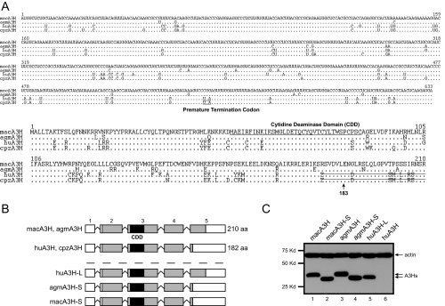
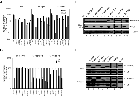
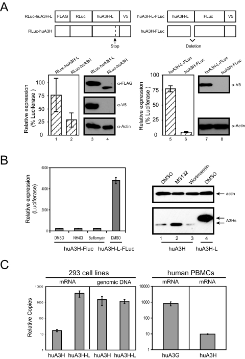
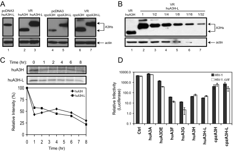
The human genome encodes seven APOBEC3 (A3) cytidine deaminases with potential antiretroviral activity: A3A, A3B, A3C, A3DE, A3F, A3G, and A3H. A3G was the first identified to block replication of human immunodeficiency virus type 1 (HIV-1) and many other retroviruses. A3F, A3B, and A3DE were shown later to have similar activities. HIV-1 produces a protein called Vif that is able to neutralize the antiretroviral activities of A3DE, A3F, and A3G, but not A3B. Only the antiretroviral activity of A3H remains to be defined due to its poor expression in cell culture. Here, we studied the mechanism impairing A3H expression. When primate A3H sequences were compared, a premature termination codon was identified on the fifth exon of the human and chimpanzee A3H genes, which significantly decreased their protein expression. It causes a 29-residue deletion from the C terminus, and this truncation did not reduce human A3H protein stability. However, the mRNA levels of the truncated gene were significantly decreased. Human A3H protein expression could be restored to a normal level either by repairing this truncation or through expression from a vector containing an intron from human cytomegalovirus. Once expression was optimized, human A3H could reduce HIV-1 infectivity up to 150-fold. Importantly, HIV-1 Vif failed to neutralize A3H activity. Nevertheless, extensive sequence analysis could not detect any significant levels of G-to-A mutation in the HIV-1 genome by human A3H. Thus, A3H inhibits HIV-1 replication potently by a cytidine deamination-independent mechanism, and optimizing A3H expression in vivo should represent a novel therapeutic strategy for HIV-1 treatment.


Figures

FIGURE 1.
FIGURE 1.
Primate A3H proteins. A, RNA and amino acid sequence alignment of primate A3H proteins. The macA3H sequence is listed on the top. Dots indicate identical sequence, and residues different from this sequence are directly indicated. The PTC in human and chimpanzee RNAs is underlined and indicated. The CDD is underlined in the amino acid sequence, and the truncated sequences are marked by strikethrough. B, schematic representation of the primate A3H exon structure. Three mutant A3H genes including huA3H-L, agmA3H-S, and macA3H-S were generated. aa, amino acids. C, expression of primate A3H genes. The cDNAs of these genes were cloned into pcDNA3.1 with a V5 tag at their 3′-end, and they were transiently expressed in 293T cells. Protein expression was determined by Western blotting with antibodies against V5 and cellular protein actin.
FIGURE 2.
FIGURE 2.
Primate A3H antiretroviral activity. A, anti-HIV-1 and SIV activity. The pcDNA3 vectors expressing huA3H-L, agmA3H, agmA3H-S, macA3H, macA3H-S, or huA3G were cotransfected with wild-type (WT) or vif-deficient HIV-1 or SIV luciferase reporter proviral clone into 293T cells at a 5:1 ratio. Equal amounts of viruses were used to infect GHOST-R3/X4/R5 cells. Viral infectivity is presented as a relative value, where the infectivity of vif-deficient virus produced in the presence of pcDNA3.1 as a control (Ctrl) is set as 100. B, package of A3H proteins in HIV-1 virions. Wild-type or vif-deficient HIV-1 virions were produced from 293T cells in the presence of huA3H-L, agmA3H-S, macA3H-S, agmA3H, macA3H, or huA3G. Virions were purified through a 20% sucrose cushion via ultracentrifugation. Viral pellets were then lysed and analyzed by Western blotting with antibodies against V5 and HIV-1 Vif and p24Gag. C, susceptibility to Vif. Primate A3H-FLuc fusion proteins were coexpressed with HIV-1 Vif, SIVagm Vif, or SIVmac Vif. Cells were then lysed, and cellular luciferase activity was determined. Data are presented as a relative value, where its parallel control transfected with a Vif deletion vector (pNL-A1ΔVif) is set as 100. D, interactions of HIV-1 Vif with primate A3H proteins. A3 proteins were C-terminally fused to a HA/FLAG tag and then coexpressed in 293T cells with HIV-1 Vif. Proteins were pulled down by Sepharose beads conjugated with anti-FLAG antibody and determined by Western blotting with anti-HA antibody for A3 proteins and anti-Vif antibody. Error bars in A and C represent S.D. in at least three independent experiments. huA2, human APOBEC2.
FIGURE 3.
FIGURE 3.
Human A3H expression. A, comparison of huA3H and huA3H-L expressions by luciferase reporter systems. Full-length and truncated human A3H proteins were fused to RLuc or FLuc at the N or C terminus, respectively. Proteins were expressed in 293T cells, and cellular luciferase activities were determined. The same cell lysates were also subject to Western blotting with antibodies against FLAG, V5, or actin. B, rescue of huA3H expression by various inhibitors. 293T cells were transfected with either FLuc fusion or non-fusion expression vectors and then treated with 40 mm NH4Cl, 0.4 μm bafilomycin, 20 μm MG132, or 100 nm wortmannin for 16 h. Human A3H protein expression was determined by measuring cellular luciferase activity or by Western blotting. DMSO, Me2SO. C, quantitation of A3H genes in vivo. Two 293 cell-derived cell lines stably expressing huA3H-FLuc or huA3H-L-FLuc were created by G418 selection. The levels of A3H mRNA and genomic DNA in these cell lines were determined by TaqMan® real-time PCR. In addition, the mRNA levels of A3G and A3H in human PBMCs were determined by SYBR Green® real-time PCR. mRNA levels were normalized to hypoxanthine-guanine phosphoribosyltransferase mRNA, and DNA levels were normalized to the amounts of DNA input. Error bars represent S.D. in at least three independent experiments.
FIGURE 4.
FIGURE 4.
Wild-type human and chimpanzee A3H anti-HIV-1 activity. A, stable expression of the wild-type huA3H and cpzA3H genes from the VR vector. The huA3H, huA3H-L, cpzA3H, and cpzA3H-L genes were cloned into the VR vector containing a C-terminal HA tag. Their expressions were then compared with those from the pcDNA3.1 vector by Western blotting after transfection into 293T cells. B, comparison of huA3H and huA3H-L expressions from the VR vector. 293T cells were transfected with the VR vector expressing huA3H or huA3H-L, and cell lysates were prepared. The huA3H-L cell lysates were serially diluted, and the levels of huA3H-L in these samples were then compared with the levels of undiluted huA3H sample by Western blotting. C, pulse-chase radiolabeling of human A3H protein. 293T cells were transiently transfected with the VR vector expressing huA3H or huA3H-L protein. Cells were pulse-labeled for 30 min and then incubated for the indicated time points. After immunoprecipitation of lysates with anti-HA antibody, proteins were separated by SDS-PAGE, and the gels were subjected to autoradiography. The relative intensity of the bands was quantified, normalized to 100, and graphed. D, anti-HIV activity. The VR vectors expressing huA3A, huA3DE, huA3F, huA3G, huA3H, huA3H-L, cpzA3H, and cpzA3H-L were cotransfected with either wild-type or vif-deficient HIV-1 luciferase reporter proviral clone into 293T cells at a 1:9 ratio (0.6 μg of the A3H expression vector plus 5.4 μg of the HIV-1 proviral vector). HIV-1 infectivity was determined as before. Error bars represent S.D. in at least three independent experiments. Ctrl, control.
FIGURE 5.
FIGURE 5.
G-to-A hypermutations in the HIV-1 genome. Wild-type (WT) or vif-deficient HIV-1 was produced in the presence of various A3 proteins. huA3H, hua3H-L, and huA3G were expressed from the VR vector. The other proteins were expressed from pcDNA3.1, which was also used as the control vector (Ctrl). These viruses were used to infect GHOST-R3/X4/R5 cells. 8 h later, cellular DNAs were extracted from infected cells, and viral DNAs from nucleotides 5707 to 6066 were amplified by PCR. After cloning into TA cloning vector, viral DNAs were sequenced. The type of mutations is summarized in tabular forms, where the original HIV-1 sequence is given at left, and the new sequence or positions from the mutated cytosine are given across the top. N at the lower right of each box indicates the total numbers of bases sequenced.

References

    1. Holmes, R. K., Malim, M. H., and Bishop, K. N. (2007) Trends Biochem. Sci. 32 118-128 - PubMed
    1. Bishop, K. N., Holmes, R. K., Sheehy, A. M., Davidson, N. O., Cho, S. J., and Malim, M. H. (2004) Curr. Biol. 14 1392-1396 - PubMed
    1. Dang, Y., Wang, X., Esselman, W. J., and Zheng, Y.-H. (2006) J. Virol. 80 10522-10533 - PMC - PubMed
    1. Doehle, B. P., Schafer, A., and Cullen, B. R. (2005) Virology 339 281-288 - PubMed
    1. Liddament, M. T., Brown, W. L., Schumacher, A. J., and Harris, R. S. (2004) Curr. Biol. 14 1385-1391 - PubMed

Publication types

LinkOut - more resources