. 2008 May;28(10):3139-50.
doi: 10.1128/MCB.01469-07. Epub 2008 Feb 25.

Generation and activation of multiple dimeric transcription factors within the NF-kappaB signaling system

Affiliations

Generation and activation of multiple dimeric transcription factors within the NF-kappaB signaling system

Soumen Basak et al. Mol Cell Biol. 2008 May.

Abstract

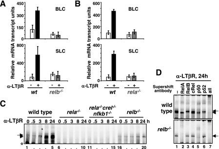
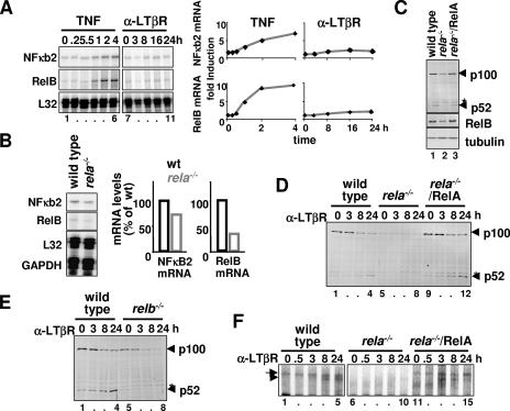
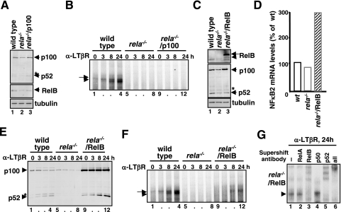
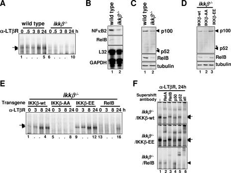
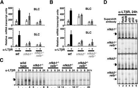
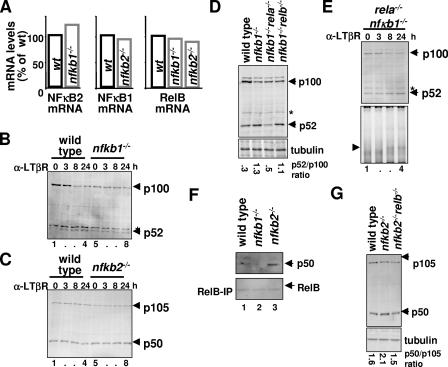
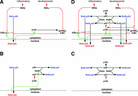
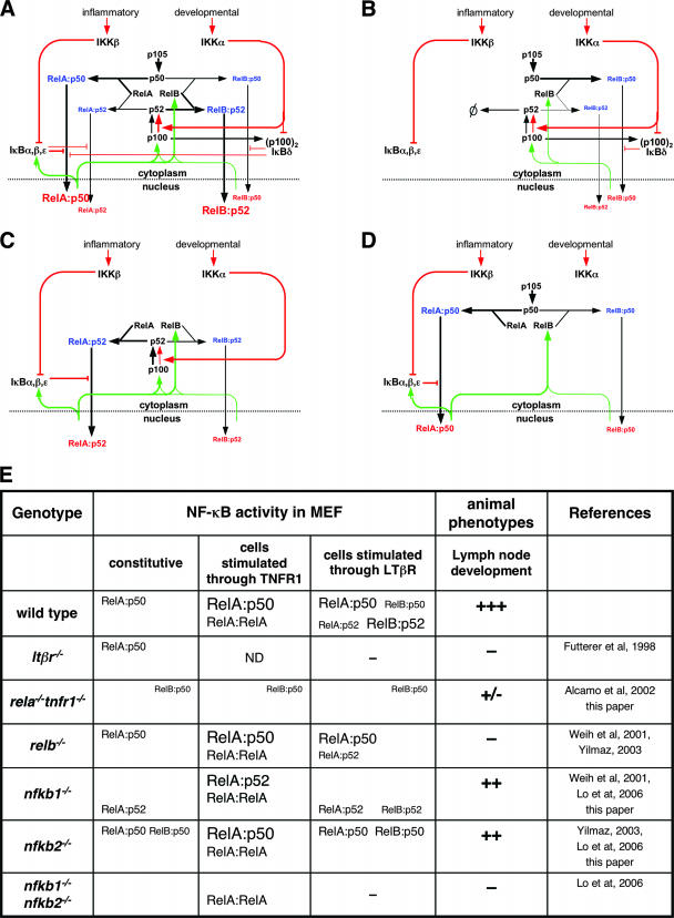
The NF-kappaB signaling pathway regulates the activity of multiple dimeric transcription factors that are generated from five distinct monomers. The availabilities of specific dimers are regulated during cell differentiation and organ development and determine the cell's responsiveness to inflammatory or developmental signals. An altered dimer distribution is a hallmark of many chronic diseases. Here, we reveal that the cellular processes that generate different NF-kappaB dimers are highly connected through multiple cross-regulatory mechanisms. First, we find that steady-state expression of RelB is regulated by the canonical pathway and constitutive RelA activity. Indeed, synthesis control of RelB is the major determinant of noncanonical NF-kappaB dimer activation. Second, processing, not synthesis, of p100 and p105 is mechanistically linked via competitive dimerization with a limited pool of RelA and RelB. This homeostatic cross-regulatory mechanism determines the availability of the p50- and p52-containing dimers and also of the noncanonical IkappaB p100. Our results inform a wiring diagram to delineate NF-kappaB dimer formation that emphasizes that inflammatory and developmental signaling cannot be considered separately but are highly interconnected.


Figures

FIG. 1.
FIG. 1.
Requirement of RelA for noncanonical NF-κB signaling induced by LTβR. (A and B) BLC and SLC mRNA levels in resting cells (open bars) and in response to LTβR stimulation as measured by real-time Q-PCR) in early-passage fibroblasts of the indicated genotypes. mRNA levels relative to the GAPDH control are plotted. (C) NF-κB DNA binding activity in MEF lines of the indicated genotypes during LTβR signaling. Specific κB-DNA binding complexes are indicated by arrows (RelA dimers) and arrowheads (RelB dimers). The asterisk denotes a nonspecific DNA-protein complex. (D) The composition of NF-κB DNA binding activity induced upon LTβR stimulation in wild-type or relb−/− MEFs was examined by supershift analysis using the indicated antibodies. At 24 h poststimulation RelA-p50 and RelB-p52 dimers appear as the major NF-κB DNA binding activity in the nucleus, with accompanying RelA-p52 and RelB-p50 dimers as minor DNA binding activity. α, anti.
FIG. 2.
FIG. 2.
RelA is required for LTβR signaling and controls steady-state RelB expression. (A) RPA to examine NF-κB2 and RelB mRNA levels in wild-type MEFs stimulated with TNF or LTβR agonist (left panel). Relative induction levels of NF-κB2 and RelB mRNA in response to TNFR or LTβR stimulation are plotted after normalizing to L32 mRNA (right panel). (B) Steady-state level of NF-κB2 and RelB mRNA in wild-type or rela−/− MEFs was examined by RPA (left panel). Respective mRNA levels were quantified and normalized to L32 and GAPDH mRNA and plotted (right panel). (C) Immunoblotting for p100 and RelB in wild-type, rela−/−, or rela−/− MEFs reconstituted with a retroviral RelA transgene. Presence of a nonspecific band is denoted with an asterisk. (D and E) Immunoblotting to examine p100 processing into p52 during LTβR signaling in MEFs of the indicated genotypes. (F) NF-κB-DNA binding activities induced upon LTβR stimulation in rela−/− MEFs that express the RelA transgene were analyzed by EMSA. α, anti.
FIG. 3.
FIG. 3.
Constitutive expression of RelB is sufficient to restore LTβR-induced RelB activation in rela−/− MEFs. (A and C) Immunoblotting to reveal p100 (A) or RelB (C) expression from transgene in rela−/− MEFs. (B and F) EMSA to examine NF-κB DNA binding activity in response to LTβR stimulation in rela−/− MEFs that expresses p100 (B) or RelB (F) from transgene. (D) Steady-state level of NF-κB2 mRNA in rela−/− MEFs in the absence or presence of the retroviral RelB transgene was analyzed by RPA. (E) Immunoblotting to examine processing of p100 and generation of p52 upon LTβR stimulation in rela−/− MEFs in the absence or presence of the retroviral RelB transgene. (G) Supershift analysis to examine the composition NF-κB DNA binding activity induced upon LTβR stimulation in rela−/− MEFs that expresses RelB transgene. α, anti.
FIG. 4.
FIG. 4.
Constitutive IKKβ activity determines LTβR-induced activation of RelB/NF-κB. (A) NF-κB DNA binding activities induced upon LTβR stimulation in MEFs of the indicated genotypes were analyzed by EMSA. (B) Steady-state levels of NF-κB2 and RelB mRNA in wild-type and ikkβ−/− MEFs was examined by RPA. (C and D) Immunoblotting to reveal the steady-state level of p100 and RelB protein in MEFs of the indicated genotypes. (E) NF-κB DNA binding activity induced by LTβR stimulation in ikkβ−/− MEFs expressing transgenic wild-type (lanes 1 to 4), inactive (lanes 5 to 8), or constitutively active forms (lanes 9 to 12) of IKKβ or ectopic RelB protein (lanes 13 to 16). (F) Supershift analysis to examine the composition of NF-κB DNA binding activity induced upon LTβR stimulation in the ikkβ−/− MEFs reconstituted with the indicated retroviral transgene. α, anti.
FIG. 5.
FIG. 5.
Requirement of both nfkb1 and nfkb2 gene products for induction of RelB target genes and NF-κB/RelB DNA binding activity in response to LTβR signaling. (A and B) Expression of BLC and SLC genes upon LTβR stimulation in primary MEFs of the indicated genotypes. (C) NF-κB DNA binding activities induced by LTβR stimulation in primary MEFs of the indicated genotypes were examined by EMSA. The presence of RelB in single nfkb1−/− and nfkb2−/− knockout cells perturbs NF-κB RelA activation. (D) Supershift analysis to reveal the composition of NF-κB DNA binding activity induced upon LTβR stimulation in the MEFs of the indicated genotypes. α, anti.
FIG. 6.
FIG. 6.
Cross-regulation between p105/p50 and p100/p52 at the level of protein processing. (A) Steady-state levels of RelB, NF-κB2, and NF-κB1 mRNAs were measured in nfkb1−/− and nfkb2−/− primary MEFs by RPA and compared with wild-type MEFs in a bar diagram. (B) Immunoblotting to reveal constitutive and LTβR-stimulated processing of p100 to p52 in wild-type and nfkb1−/− primary MEFs. (C) The levels of p105 and p50 proteins in resting and LTβR-stimulated wild-type and nfkb2−/− MEFs were examined by immunoblot analysis. (D) Homeostatic level of p52 and p100 in primary MEFs of the indicated genotypes were examined by immunoblot analysis. Band intensities were quantified using ImageQuant software, normalized to tubulin, and expressed as a ratio of processed product to precursor. (E) Immunoblot (top) and EMSA (bottom) analysis to reveal p52 accumulation and RelB-p52 dimer activation, respectively, in nfkb1−/− rela−/− MEFs in response to LTβR stimulation. (F) The levels of p50 (top) associated with RelB (bottom) in wild-type and nfkb2−/− MEFs were examined by immunoblot analysis of the RelB immunoprecipitate obtained from the indicated cell extracts. (G) Homeostatic level of p50 and p105 in primary MEFs of the indicated genotypes were examined by immunoblot analysis, and the ratios of processed product to precursor were quantified. α, anti.
FIG. 7.
FIG. 7.
Wiring diagrams to depict functional relationships within the NF-κB signaling system. NF-κB monomers are shown in black, cytoplasmic dimers are in blue, and nuclear dimers are in red. Constitutive processes are shown in black, regulated processes are in red, and feedback processes are in green. (A) A wiring diagram of the regulation of the NF-κB/RelA-p50 dimer in response to inflammatory or developmental stimuli. A mathematical model of these functional connections has been constructed and shown to recapitulate signaling cross talk between these pathways in the control of RelA-p50 activity (2). (B) A wiring diagram to summarize our findings (Fig. 2 and 3) that steady-state RelB synthesis and the availability of latent RelB dimers are dependent on constitutive RelA-p50 activity. In Fig. 4, we also showed that constitutive RelA-p50 activity is controlled by constitutive IKKβ activity. (C) A wiring diagram to describe the molecular competition between p50 and p52 for their dimerization partners RelA and RelB. This competition determines the rate of p50 and p52 generation via processing from their p105 and p100 precursors. Thus, p105/p50 and p100/p52 processing is interdependent through competition for common interaction partners. (D) A wiring diagram of the NF-κB signaling system that accounts for the generation of four NF-κB dimers that are detected in response to LTβR signaling, namely, RelA-p50, RelA-p52, RelB-p50, and RelB-p52. This diagram is based on previously established connectivity (A) and incorporates the insights from the current study summarized in panels B and C. The NF-κB signaling system receives signals from both inflammatory and developmental cues through IKK2/IKKβ and IKK1/IKKα kinases, respectively, to activate distinct NF-κB dimers. For the sake of clarity, we have depicted only signal-responsive protein complexes, omitting dimers such as p50-p50, p105-p50, p105-RelA, p100-p52, or p100-RelB that do not change in abundance during LTβR induced signaling.
FIG. 8.
FIG. 8.
(A) A modified version of the wiring diagram that now indicates the preferred biochemical reactions with bold lines and lesser reactions with thin lines to more quantitatively mirror the NF-κB signaling system in wild-type cells that responds to both inflammatory and developmental signaling. (B to D) Wiring diagrams that describe mutant NF-κB signaling systems in rela−/−, nfkb1−/−, or nfkb2−/− cells, respectively. (E) A comprehensive description of the NF-κB dimers that are activated during signaling through TNFR1 or LTβR in MEFs of the indicated genotypes. Relative DNA binding activities of a given NF-κB dimer under various conditions are represented with different font sizes. ND, not determined. Observed lymph node phenotypes of various NF-κB gene knockout mice are shown: +++, normal development; −, severe defect in lymph node development; ++ and +/−, intermediate phenotypes.

References

    1. Alcamo, E., N. Hacohen, L. C. Schulte, P. D. Rennert, R. O. Hynes, and D. Baltimore. 2002. Requirement for the NF-κB family member RelA in the development of secondary lymphoid organs. J. Exp. Med. 195233-244. - PMC - PubMed
    1. Basak, S., H. Kim, J. D. Kearns, V. Tergaonkar, E. O'Dea, S. L. Werner, C. A. Benedict, C. F. Ware, G. Ghosh, I. M. Verma, and A. Hoffmann. 2007. A fourth IκB protein within the NF-κB signaling module. Cell 128369-381. - PMC - PubMed
    1. Beinke, S., and S. C. Ley. 2004. Functions of NF-κB1 and NF-κB2 in immune cell biology. Biochem. J. 382393-409. - PMC - PubMed
    1. Bonizzi, G., M. Bebien, D. C. Otero, K. E. Johnson-Vroom, Y. Cao, D. Vu, A. G. Jegga, B. J. Aronow, G. Ghosh, R. C. Rickert, and M. Karin. 2004. Activation of IKKαtarget genes depends on recognition of specific κB binding sites by RelB:p52 dimers. EMBO J. 234202-4210. - PMC - PubMed
    1. Bonizzi, G., and M. Karin. 2004. The two NF-kappaB activation pathways and their role in innate and adaptive immunity. Trends Immunol. 25280-288. - PubMed

Publication types

MeSH terms