. 2008 Mar 4;105(9):3533-8.
doi: 10.1073/pnas.0710951105. Epub 2008 Feb 25.

PD-1 deficiency reveals various tissue-specific autoimmunity by H-2b and dose-dependent requirement of H-2g7 for diabetes in NOD mice

Affiliations

PD-1 deficiency reveals various tissue-specific autoimmunity by H-2b and dose-dependent requirement of H-2g7 for diabetes in NOD mice

Taku Yoshida et al. Proc Natl Acad Sci U S A. .

Abstract

Although many autoimmune diseases are associated with particular HLA/H-2 haplotypes, the mechanisms through which specific HLA/H-2 haplotypes afford autoimmune susceptibility remain enigmatic. Here, we analyzed the effects of the diabetes-associated (H-2(g7)) and an antidiabetogenic (H-2(b)) H-2 haplotypes in NOD mice deficient for programmed cell death-1 (PD-1, Pdcd1), a unique model of type 1 diabetes that confers complete penetrance and rapid onset of the disease. NOD-H2(b/b)Pdcd1(-/-) mice were completely protected from diabetes, confirming that H-2(g7) is indispensable for diabetes even in the absence of PD-1. However, NOD-H2(b/b)Pdcd1(-/-) mice developed autoimmune inflammation in multiple tissues including peripheral nerves, stomachs, and exocrine tissues, demonstrating that autoreactive T cells are generated in the context of H-2(b). These autoreactive T cells damaged target tissues only in the absence of PD-1, confirming that PD-1 deficiency unravels the hidden autoimmune susceptibility of the strain by reducing the threshold of T cell activation. Transfer experiments revealed that CD4 T cells are the effector cells of neuritis, and nerve-infiltrating CD4 T cells are strongly deviated toward Th1. Interestingly, neuritogenic T cells were also generated in the context of H-2(g7), in sharp contrast to the strict requirement of H-2(g7) for diabetes. In addition, 60% of the NOD-H2(b/g7)Pdcd1(-/-) mice developed diabetes, suggesting that H-2(b) does not dominantly suppress diabetes and that H-2(g7) induces diabetes in a dose-dependent fashion.


Conflict of interest statement

The authors declare no conflict of interest.

Figures

Fig. 1.
Fig. 1.
Complete protection of NOD-Pdcd1−/− mice from T1DM by H-2b/b but not H-2b/g7. (a and b) Incidence of T1DM in female (a) and male (b) mice is shown for NOD-H2g7/g7Pdcd1+/+ (open circles), NOD-H2b/g7Pdcd1+/+ (open triangles), NOD-H2b/bPdcd1+/+ (open squares), NOD-H2g7/g7Pdcd1−/− (closed circles), NOD-H2b/g7Pdcd1−/− (closed triangles), and NOD-H2b/bPdcd1−/− (closed squares) mice (n = 11 ≈ 28). (c) Representative H&E staining of islets is shown for female NOD-Pdcd1+/+ (Top) and NOD-Pdcd1−/− (Middle) mice with H-2g7/g7 (Left), H-2b/g7 (Center), and H-2b/b (Right) haplotypes. Sections were also stained with Abs against CD4 (red), CD8 (blue), and PD-L1 (green) (Bottom). (Original magnification, ×400.)
Fig. 2.
Fig. 2.
Spontaneous development of peripheral polyneuropathy by NOD-H2b/g7Pdcd1−/− and NOD-H2b/bPdcd1−/− mice. (a) Incidence of polyneuropathy is shown for female NOD-H2g7/g7Pdcd1+/+ (open circles), NOD-H2b/g7Pdcd1+/+ (open triangles), NOD-H2b/bPdcd1+/+ (open squares), NOD-H2g7/g7Pdcd1−/− (closed circles), NOD-H2b/g7Pdcd1−/− (closed triangles), and NOD-H2b/bPdcd1−/− (closed squares) mice (n = 11 ≈ 28). NOD-H2g7/g7Pdcd1−/− mice were killed at 10 weeks of age because of the severe T1DM. (b) A neuropathic mouse with severe paralysis of the limbs is shown. (c) Luxol fast blue staining of the sciatic nerve (Left) and H&E stainings of the sciatic nerve (Center) and dorsal root ganglion (Right) are shown. (d) Severity of neuropathy in male (closed circles) and female (open circles) mice is shown for the indicated genotypes. (e) Sciatic nerves from NOD-H2b/bPdcd1+/+ (Upper) and NOD-H2b/bPdcd1−/− (Lower) mice were stained with Abs against CD4 (green), CD8 (blue), and IgM (red) (Left), PD-L1 (green), CD4 (red), and CD8 (blue) (Center), and PD-L1 (green) and CD11c (red) (Right). (Original magnification: c and e, ×200.)
Fig. 3.
Fig. 3.
CD4 T cells are the effector cells of the polyneuropathy. (a–c) Representative H&E staining of sciatic nerve from C57BL/6-Rag2−/− mice that had received transfers of unfractionated splenocytes (a), CD4 T cells (b), or CD4-depleted splenocytes (c) prepared from the spleen of neuropathic NOD-H2b/bPdcd1−/− mice. (d) The severity of polyneuropathy is shown for each recipient mouse. (e) B cells are dispensable for the development of polyneuropathy. Representative H&E staining of NOD-H2b/bPdcd1−/−μMT mouse is shown. (f–i) NOD-H2b/bPdcd1−/− mice produce autoAbs against myelin sheath. (f) Sciatic nerve from NOD-SCID mouse was stained with serum from neuropathic NOD-H2b/bPdcd1−/− mouse. (g–i) Higher magnification of sciatic nerve stained with Luxol fast blue (g), serum (green), and anti-neuronal class III β-tubulin Ab (red) (h), or serum (green) and anti-MBP Ab (red) (i). Representative data of more than five mice are shown. (Original magnification: a–f, ×200; g–i, ×630.)
Fig. 4.
Fig. 4.
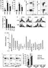
Th1-biased activation of the nerve-infiltrating T cells. (a) The fraction of CD44+, CD62L, and CD69+ cells is shown for CD4 (Left) and CD8 (Right) T cells in the spleen of 25-wk-old NOD-H2b/bPdcd1+/+ (white bars) and NOD-H2b/bPdcd1−/− (black bars) mice. (b) Memory CD4 T cells (Left) and CD8 T cells (Right) in spleen are shown for NOD-H2b/bPdcd1+/+ (Top) and NOD-H2b/bPdcd1−/− (Lower) mice. (c) Statistics of the cellular composition of the nerve-infiltrating cells are shown. (d) Expression of indicated molecules on the sciatic nerve-infiltrating cells (thick lines) and splenocytes (filled histograms) is shown for CD4 (Upper) and CD8 (Lower) T cells. (e) Increase of Th1 and inflammatory cytokines in the inflamed nerve. Relative quantities of the mRNAs for the indicated genes in the inflamed nerve in relation to control nerve are shown. Data are the mean + SEM of three mice. (f) Representative FACS profiles of INFγ- and IL-4-stained spleen cells from NOD-H2b/bPdcd1+/+ (Left) and NOD-H2b/bPdcd1−/− (Center) mice and of nerve-infiltrating cells from NOD-H2b/bPdcd1−/− mice (Right) are shown for CD4 (Upper) and CD8 (Lower) T cells. (g) Mean percentage of INFγ- (Left), IL-4- (Center), and IL-17- (Right) producing CD4 and CD8 T cells is shown. Data are the mean + SEM. Representative data of more than three experiments are shown. *, P < 0.05; **, P < 0.01.
Fig. 5.
Fig. 5.
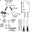
Neuritogenic T cells can be generated in the context of either H-2b or H-2g7. (a) Total spleen cells from neuropathic NOD-H2b/g7Pdcd1−/− mice were injected into C57BL/6-Rag2−/− and NOD-SCID mice (Left). The severity of polyneuropathy is shown for each recipient mouse (Right). (b) Development of neuritis in the absence of H-2b. The incidence of severe (black) and moderate (gray) neuritis is shown for female nondiabetic [(NOD-H2g7/g7Pdcd1−/− × C57BL/6-H2b/bPdcd1−/−)F1 × NOD-H2g7/g7Pdcd1−/−]BC1 mice with H2g7/g7 (Left, n = 12) and H2b/g7 (Right, n = 41) at 24–30 weeks of age.

References

    1. Anderson MS, Bluestone JA. The NOD mouse: A model of immune dysregulation. Annu Rev Immunol. 2005;23:447–485. - PubMed
    1. Wicker LS, Todd JA, Peterson LB. Genetic control of autoimmune diabetes in the NOD mouse. Annu Rev Immunol. 1995;13:179–200. - PubMed
    1. Wicker LS, et al. Autoimmune syndromes in major histocompatibility complex (MHC) congenic strains of non-obese diabetic (NOD) mice. The NOD MHC is dominant for insulitis and cyclophosphamide-induced diabetes. J Exp Med. 1992;176:67–77. - PMC - PubMed
    1. Wicker LS, Miller BJ, Fischer PA, Pressey A, Peterson LB. Genetic control of diabetes and insulitis in the non-obese diabetic mouse. Pedigree analysis of a diabetic H-2nod/b heterozygote. J Immunol. 1989;142:781–784. - PubMed
    1. Carrasco-Marin E, Shimizu J, Kanagawa O, Unanue ER. The class II MHC I-Ag7 molecules from non-obese diabetic mice are poor peptide binders. J Immunol. 1996;156:450–458. - PubMed

Publication types