

Spinal epidural abscess -- a rare complication of inflammatory bowel disease
- PMID: 18299737
- PMCID: PMC2659139
- DOI: 10.1155/2008/893757
Spinal epidural abscess -- a rare complication of inflammatory bowel disease
Abstract
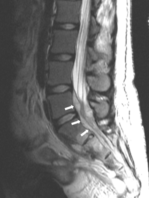
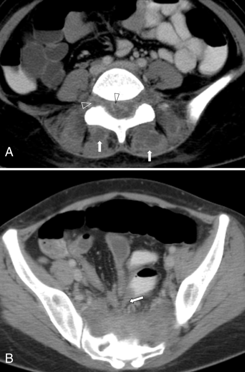
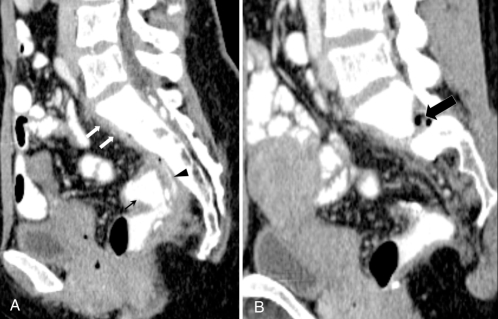
Spinal epidural abscess is an uncommon but highly morbid illness. While it usually afflicts older, immunocompromised patients, this condition has been reported as a result of intestinal perforation in the setting of inflammatory bowel disease. Two cases of spinal epidural abscess in patients with inflammatory bowel disease are reported: one in a patient with Crohn's disease and one in a patient with ulcerative colitis after restorative proctocolectomy.
L’abcès spinal épidural est une maladie rare mais extrêmement morbide. Bien qu’il affecte généralement des patients plus âgés et immunodéprimés, ce type d’abcès a été signalé après une perforation intestinale dans le contexte de la maladie inflammatoire de l’intestin. Deux cas d’abcès spinal épidural chez des patients atteints de maladie inflammatoire de l’intestin sont décrits ici. L’un chez un patient atteint de la maladie de Crohn et l’autre, chez un patient atteint de colite ulcéreuse ayant subi une proctocolectomie correctrice.
Figures
References
-
- Bell SJ, Williams AB, Wiesel P, Wilkinson K, Cohen RC, Kamm MA. The clinical course of fistulating Crohn’s disease. Aliment Pharmacol Ther. 2003;17:1145–51. - PubMed
-
- Schwartz DA, Loftus EV, Jr, Tremaine WJ, et al. The natural history of fistulizing Crohn’s disease in Olmsted County, Minnesota. Gastroenterology. 2002;122:875–80. - PubMed
-
- Ben-Ami H, Ginesin Y, Behar DM, Fischer D, Edoute Y, Lavy A. Diagnosis and treatment of urinary tract complications in Crohn’s disease: An experience over 15 years. Can J Gastroenterol. 2002;16:225–9. - PubMed
-
- Yamamoto T, Keighley MR. Enterovesical fistulas complicating Crohn’s disease: Clinicopathological features and management. Int J Colorectal Dis. 2000;15:211–5. - PubMed
-
- Wulfeck D, Williams T, Amin A, Huang TY. Crohn’s disease with unusual enterouterine fistula in pregnancy. J Ky Med Assoc. 1994;92:267–9. - PubMed
Publication types
MeSH terms
LinkOut - more resources
Full Text Sources
Medical