

Immunopathological aspects of age-related macular degeneration
- PMID: 18299834
- PMCID: PMC2441602
- DOI: 10.1007/s00281-008-0112-9
Immunopathological aspects of age-related macular degeneration
Abstract
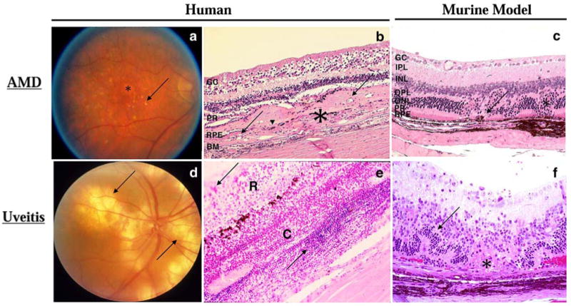
Age-related macular degeneration (AMD) represents a leading cause of blindness worldwide. While the clinical and histopathological aspects of AMD are well characterized, its etiology and pathogenesis remain unclear. Recent findings suggest a role for immunologic processes in AMD pathogenesis, including the age-related generation of extracellular deposits inside the Brusch membrane and beneath the retinal pigment epithelium, recruitment of macrophages for clearance of these deposits, complement activation, recruitment of tissue-destructive macrophages, microglial activation and accumulation, and proinflammatory effects of chronic inflammation by Chlamydia pneumoniae. This review discusses the evidence for the role of inflammation in human AMD and in animal models of AMD.
Figures
References
-
- Klein R, Peto T, Bird A, Vannewkirk MR. The epidemiology of age-related macular degeneration. Am J Ophthalmol. 2004;137:486–495. - PubMed
-
- Friedman DS, O'Colmain BJ, Munoz B, Tomany SC, McCarty C, de Jong PT, Nemesure B, Mitchell P, Kempen J. Prevalence of age-related macular degeneration in the United States. Arch Ophthalmol. 2004;122:564–572. - PubMed
-
- Klein R, Klein BE, Linton KL. Prevalence of age-related maculopathy. The Beaver Dam Eye Study. Ophthalmology. 1992;99:933–943. - PubMed
-
- Klein R, Klein BE, Jensen SC, Meuer SM. The five-year incidence and progression of age-related maculopathy: the Beaver Dam Eye Study. Ophthalmology. 1997;104:7–21. - PubMed
-
- Ambati J, Ambati BK, Yoo SH, Ianchulev S, Adamis AP. Age-related macular degeneration: etiology, pathogenesis, and therapeutic strategies. Surv Ophthalmol. 2003;48:257–293. - PubMed
Publication types
MeSH terms
Grants and funding
LinkOut - more resources
Full Text Sources
Other Literature Sources
Medical
