

Pseudomyxoma peritonei: is disease progression related to microbial agents? A study of bacteria, MUC2 AND MUC5AC expression in disseminated peritoneal adenomucinosis and peritoneal mucinous carcinomatosis
- PMID: 18299935
- PMCID: PMC2570966
- DOI: 10.1245/s10434-007-9778-9
Pseudomyxoma peritonei: is disease progression related to microbial agents? A study of bacteria, MUC2 AND MUC5AC expression in disseminated peritoneal adenomucinosis and peritoneal mucinous carcinomatosis
Abstract
Background and aims: Pseudomyxoma peritonei (PMP) is characterized by peritoneal tumors arising from a perforated appendiceal adenoma or adenocarcinoma, but associated entry of enteric bacteria in the peritoneum has not been considered as a cofactor. Because Gram-negative organisms can upregulate MUC2 mucin gene expression, we determined whether bacteria were detectable in PMP tissues.
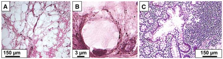
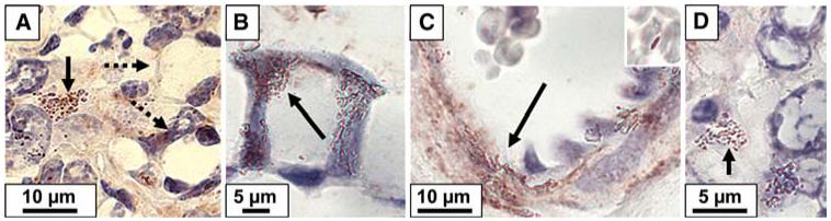
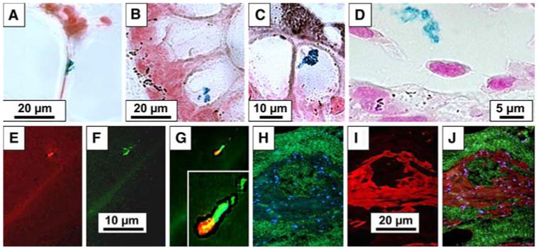
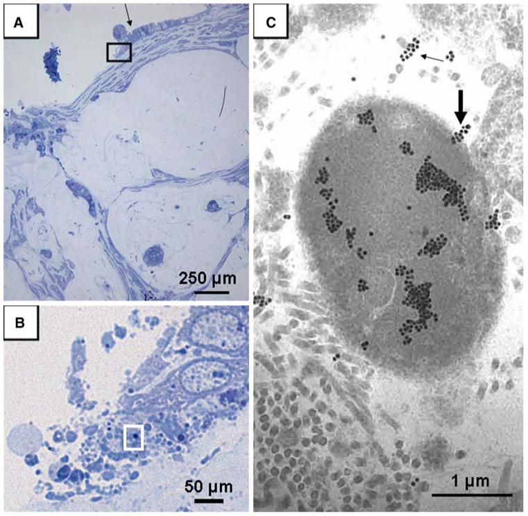
Methods: In situ hybridization was performed on resection specimens from five control subjects with noninflamed, nonperforated, non-neoplastic appendix and 16 patients with PMP [six with disseminated peritoneal adenomucinosis (DPAM) and 10 with peritoneal mucinous carcinomatosis (PMCA)]. Specific probes were designed to recognize: (1) 16S rRNA common to multiple bacteria or specific to H. pylori; (2) H. pylori cagA virulence gene; or (3) MUC2 or MUC5AC apomucins. Specimens from one patient with PMCA were examined by ultrastructural immunohistochemistry. Bacterial density and apomucin expression were determined in four histopathological compartments (epithelia, inflammatory cells, stroma, and free mucus).
Results: Enteric bacteria were detected in all specimens. Bacterial density and MUC2 expression were significantly (p < 0.05) higher in PMCA than in DPAM and controls and were highest in free mucin. MUC2 was also expressed in dysplastic epithelia and in associated inflammatory cells. MUC2 expression was significantly correlated with bacterial density.
Conclusions: Multiple enteric bacteria are present in PMP, and bacterial density and MUC2 expression is highest in the malignant form of PMP. Based on these observations, we propose that the bacteria observed in PMP may play a role in the mucinous ascites and perhaps promote carcinogenesis.
Figures
References
-
- Bradley RF, Stewart JH, Russell GB, et al. Pseudomyxoma peritonei of appendiceal origin: a clinicopathologic analysis of 101 patients uniformly treated at a single institution, with literature review. Am J Surg Pathol. 2006;30:551–9. - PubMed
-
- Mann WJ, Jr., Wagner J, Chumas J, et al. The management of pseudomyxoma peritonei. Cancer. 1990;66:1636–40. - PubMed
-
- Aho AJ, Heinonen R, Lauren P. Benign and malignant mucocele of the appendix. Histological types and prognosis. Acta Chir Scand. 1973;139:392–400. - PubMed
-
- Witkamp AJ, de Bree E, Kaag MM, van Slooten GW, van CF, Zoetmulder FA. Extensive surgical cytoreduction and intraoperative hyperthermic intraperitoneal chemotherapy in patients with pseudomyxoma peritonei. Br J Surg. 2001;88:458–63. - PubMed
-
- Galani E, Marx GM, Steer CB, et al. Pseudomyxoma peritonei: the ‘controversial’ disease. Int J Gynecol Cancer. 2003;13:413–8. - PubMed
Publication types
MeSH terms
Substances
Grants and funding
LinkOut - more resources
Full Text Sources
Medical
Miscellaneous
