. 2008 Jan;14(1):62-72.
doi: 10.1080/13550280701809084.

Platelet-derived growth factor protects neurons against gp120-mediated toxicity

Affiliations

Platelet-derived growth factor protects neurons against gp120-mediated toxicity

Fuwang Peng et al. J Neurovirol. 2008 Jan.

Abstract

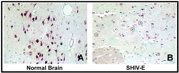
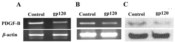
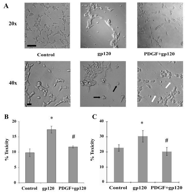
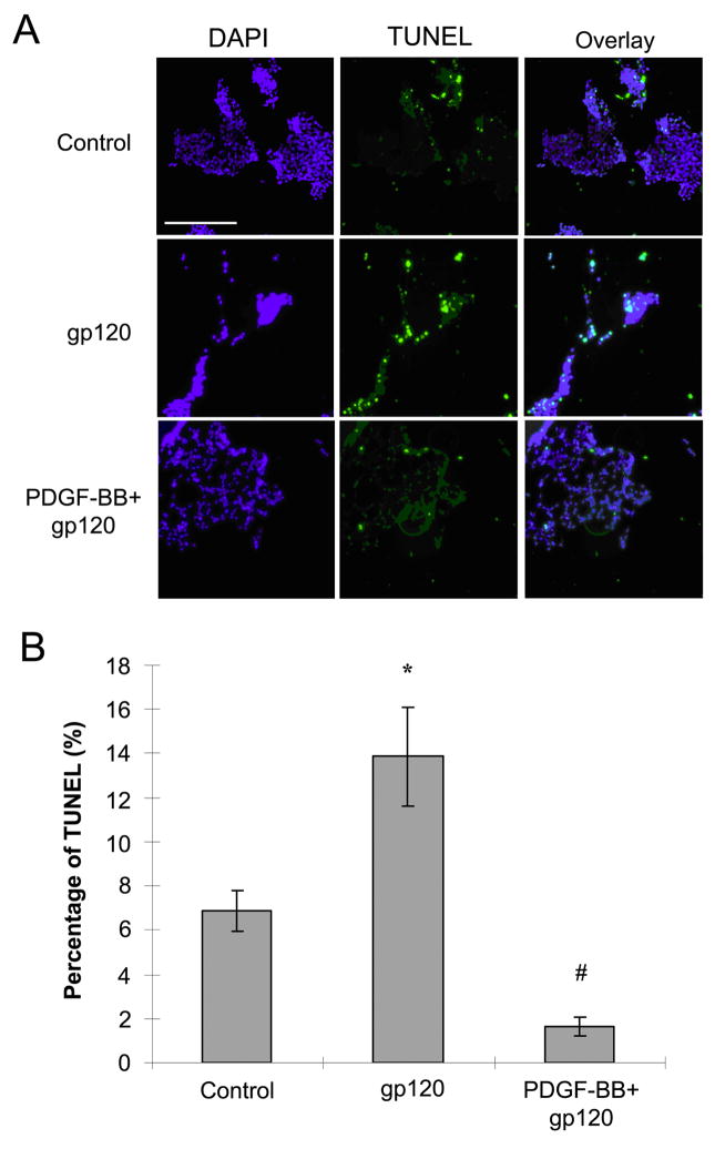
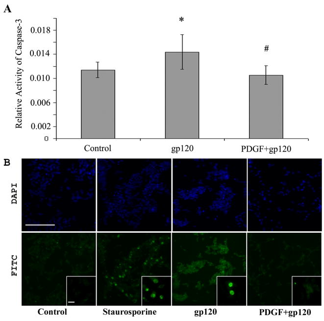
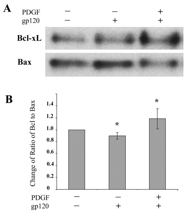
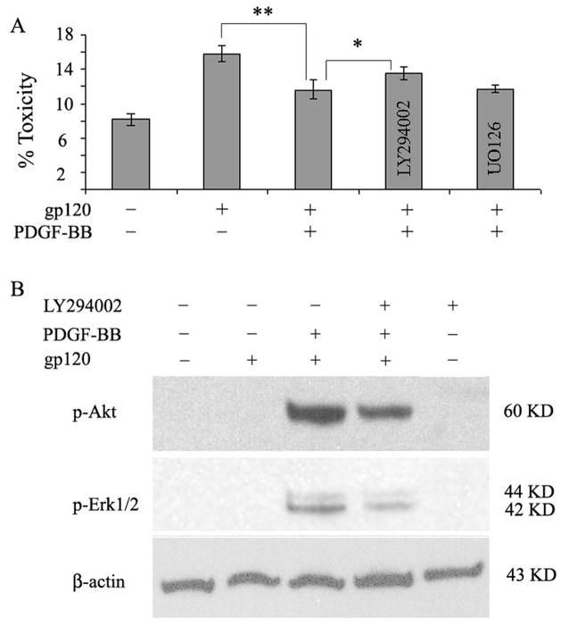
The human immunodeficiency virus (HIV)-1 envelope glycoprotein gp120 has been implicated in mediating neuronal apoptosis, a hallmark feature of HIV-associated dementia (HAD). Mitigation of the toxic effects of gp120 could thus be a potential mechanism for reducing HIV toxicity in the brain. In this study the authors hypothesized that neurotrophic factor, such as platelet-derived growth factor (PDGF), could protect the neurons against gp120-mediated apoptosis. SH-SY5Y cells treated with gp120 exhibited increased cell death when measured by lactate dehydrogenase (LDH) and deoxynucleotidyltransferase-mediated dUTP nick end labeling (TUNEL) assay, with concomitant loss of neurites and increased cell rounding. Pretreatment with PDGF-BB, however, reduced gp120-associated neurotoxicity and rescued the neurite outgrowth. Additionally, gp120-mediated activation of caspase-3 was also significantly reduced in cells pretreated with PDGF-BB. Antiapoptotic effects of PDGF-BB were also confirmed by monitoring levels of anti- and proapoptotic genes, Bcl-xL and Bax, respectively. Furthermore, PDGF-mediated protection against gp120 involved the phosphoinositide (PI) 3-kinase/Akt pathway. Taken together these findings lead us to suggest that PDGF-BB could be considered as a therapeutic agent that can mitigate gp120-mediated neurotoxicity in HAD.


Figures

Figure 1
Figure 1
Decrease of PDGF-B chain in neurons of macaque brain with SHIV-encephalitis. Representative brain sections from the basal ganglia region of an uninfected (A) and SHIV-E (B) macaque immunostained with PDGF-B chain antibody. Red color represents PDGF-B chain positive cells. Degenerated neurons are shown in circle.
Figure 2
Figure 2
Down regulation of PDGF-B chain in neurons exposed to gp120. RT-PCR analysis for PDGF-B chain was done on total RNA extracted from SH-SY5Y cells (A) or primary rat neurons (B) exposed to 200ng/ml gp120 for 24h. C) Western blot analysis of PDGF-B chain on cell lysate obtained from SH-SY5Y cells treated with or without gp-120 (200ng/ml) for 24hrs. Figure shown is a representative of three different experiments.
Figure 3
Figure 3
PDGF protects SH-SY5Y cells against gp120-mediated toxicity. A) Phase contrast microscopy of SH-SY5Y cells treated with gp120 (200ng/ml) and/or PDGF-BB (20ng/ml) for 5 days. Cells treated with gp120 alone showed rounding (as shown by black arrows) with shortened neurites. Pre-treatment of gp120-treated neurons with PDGF-BB resulted in maintenance of cellular architecture with extended neuronal processes (indicated by white arrows). Scale bars, 50 μM. Neuronal viability in the presence of gp120 and/or PDGF-BB. SH-SY5Y cells (B) and rat primary neuronal culture (C) were exposed to gp120 (200ng/ml) with or without pre-treatment with PDGF-BB (20ng/ml). Supernatant fluids and cellular extracts were collected in 24h for LDH assay. Data are presented as mean ± SEM from three independent experiments. Statistical analysis was performed using Student’s t-test. (*p < 0.01 gp120 vs control, #p<0.01 gp120 vs PDGF+gp120).
Figure 4
Figure 4
PDGF-BB mediates neuronal protection against gp120. A) SH-SY5Y cells treated with or without gp120 (200ng/ml) in the presence or absence of PDGF-BB (20ng/ml) for 18h were stained for TUNEL. Scale bar, 100 μM B) Histogram showing the percentage of TUNNEL positive SH-SY5Y cells relative to total number of neurons following various treatments. Six to eight images were randomly taken for counting positive signal in each experiment. Values are presented as mean ± SEM. (* p<0.01 gp120 vs control, #p<0.001 gp120 vs PDGF+gp120).
Figure 5
Figure 5
Neuronal apoptosis in SH-SY5Y cells exposed to gp120 with or without PDGF-BB pretreatment. Cells treated with or without gp120 IIIB in presence or absence of PDGF-BB pre-treatment were monitored for: A) caspase-3 activity assay in cell lysates and B) active caspase-3 staining by immunocytochemistry using the anti-cleaved caspase-3 antibody. The immunochemistry images showing nuclear (upper panel) and active caspase-3 staining (lower panel) were captured on Nikon inverted fluorescence microscope TE2000-E with a 20x and 100x (inset) objective (scale bar, 100 μM). Data for caspase-3 activity represents the mean ± SEM from seven independent experiments (*P<0.05 gp120 vs control, #P<0.01 gp120 vs PDGF+gp120).
Figure 6
Figure 6
Ratio of anti- and pro-apoptotic markers in PDGF-mediated neuroprotection of SH-SY5Y cells. A) Cells treated with or without gp120 in the absence or presence of PDGF-BB pre-treatment were lysed and assayed for levels of anti- and pro-apoptotic markers Bcl-xL and Bax, respectively by Western blot analysis in the same membrane. B) Densitometry scan of the ratio of band intensities of Bcl-xL/Bax from three different western blot experiments (*p<0.05, treatment vs control).
Figure 7
Figure 7
Involvement of PI3k/Akt pathway in PDGF-BB mediated protection against gp120 toxicity. A) LDH assay of SH-SY5Y cells pretreated with or without PI3K inhibitor (LY294002) or MEK1/2 inhibitor (U0126) followed by PDGF and/or gp120 exposure. Pre-treatment of cells with PI3K inhibitor resulted in significant (P value <0.05 PDGF+gp120+LY vs PDGF+gp120) reduction of PDGF-mediated protection. Pre-treatment with Erk1/2 inhibitor did not reverse the protective effect of PDGF. Data represents mean ± SEM from three independent experiments. B) Western blot analysis of lysates of phosphorylated Akt and Ek1/2 in SH-SY5Y cells pretreated with or without PI3K inhibitor (10uM LY294002) prior to PDGF and/or gp120 exposure. Blots were reprobed with antibody specific for β-actin for normalization.

Similar articles

Cited by

References

    1. Adamson DC, Dawson TM, Zink MC, Clements JE, Dawson VL. Neurovirulent simian immunodeficiency virus infection induces neuronal, endothelial, and glial apoptosis. Mol Med. 1996;2:417–428. - PMC - PubMed
    1. Bachis A, Mocchetti I. Brain-derived neurotrophic factor is neuroprotective against human immunodeficiency virus-1 envelope proteins. Ann N Y Acad Sci. 2005;1053:247–257. - PubMed
    1. Bachis A, Mocchetti I. Semisynthetic sphingoglycolipid LIGA20 is neuroprotective against human immunodeficiency virus-gp120-mediated apoptosis. J Neurosci Res. 2006;83:890–896. - PubMed
    1. Bansal AK, Mactutus CF, Nath A, Maragos W, Hauser KF, Booze RM. Neurotoxicity of HIV-1 proteins gp120 and Tat in the rat striatum. Brain Res. 2000;879:42–49. - PubMed
    1. Bell JE. The neuropathology of adult HIV infection. Rev Neurol (Paris) 1998;154:816–829. - PubMed

Publication types

MeSH terms