. 2008 Mar;9(3):310-8.
doi: 10.1038/ni1558.

A mechanism for the initiation of allergen-induced T helper type 2 responses

Affiliations

A mechanism for the initiation of allergen-induced T helper type 2 responses

Caroline L Sokol et al. Nat Immunol. 2008 Mar.

Abstract

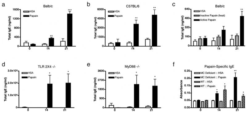
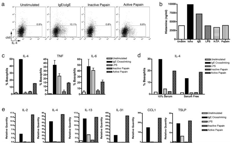
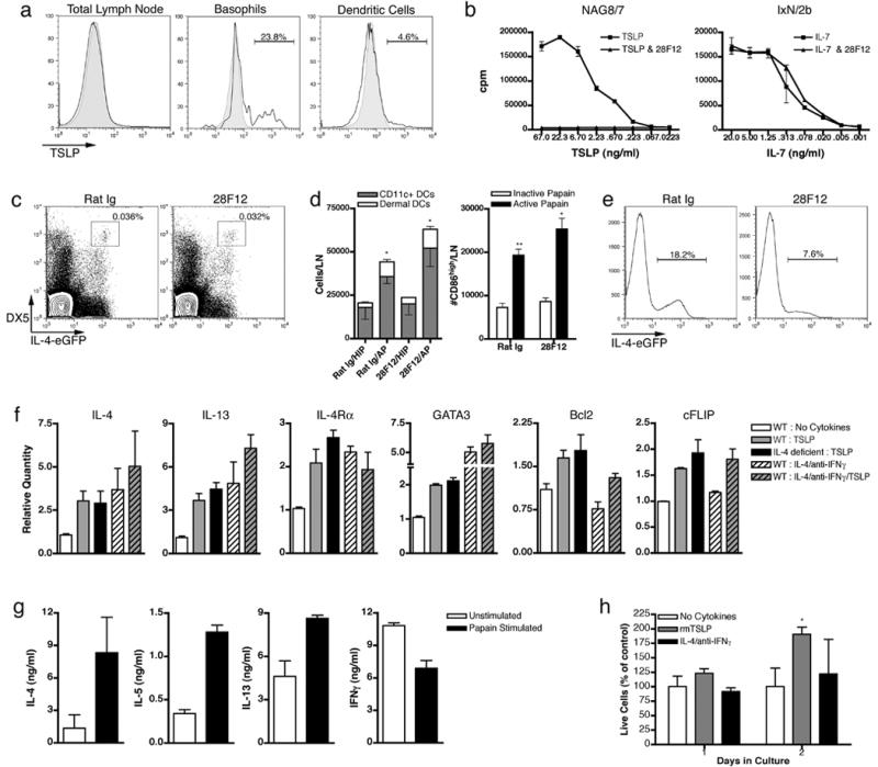
Both metazoan parasites and simple protein allergens induce T helper type 2 (TH2) immune responses, but the mechanisms by which the innate immune system senses these stimuli are unknown. In addition, the cellular source of cytokines that control TH2 differentiation in vivo has not been defined. Here we showed that basophils were activated and recruited to the draining lymph nodes specifically in response to TH2-inducing allergen challenge. Furthermore, we demonstrate that the basophil was the accessory cell type required for TH2 induction in response to protease allergens. Finally, we show that basophils were directly activated by protease allergens and produced TH2-inducing cytokines, including interleukin 4 and thymic stromal lymphopoietin, which are involved in TH2 differentiation in vivo.


Figures

Figure 1
Figure 1
Protease active papain induces IgE secretion independent of TLR2, TLR4 or MyD88 signaling. Serum IgE levels immediately before primary immunization (day 0), before secondary immunization (day 14) and at day 21 in Balb/c (a) or C57BL/6 (b) mice are shown. (c) Total IgE response to heat inactivated papain in Balb/c mice, in TLR2/4 -/- mice (d), or MyD88 -/- mice (e). (f) Antigen specific IgE response in mast cell (MC) deficient (KitW/W-v) and littermate controls (WT). For each timepoint, n=5-10 (a-c), or n=3-5 (d-f). Data are representative of at least three separate experiments, panels d and e represent data combined from separate experiments. Error bars represent s.e.m.; p values are calculated via the Student's t test, and represent comparisons to HSA immunizations at the indicated time-points; ***, p0.0001; **, p0.001, *, p0.01. If not indicated p>0.05.
Figure 2
Figure 2
The protease activity of papain has adjuvant activity and leads to Th2 cell development on day 4 post immunization. (a) 4get mice were immunized with the indicated stimuli and CD4+DX5- cells were examined for IL-4-eGFP expression. (b) 4get mice were immunized with papain or heat inactivated papain and Th2 differentiation was examined on indicated days post immunization. (c) CD4+ cells from DO11.10x4get mice were transferred into Balb/c mice, immunized as indicated and KJ1.26+CD4+ T cells were examined for IL-4-eGFP expression on day 4 post immunization, except for the OVA in Alum condition, which was examined at day 11 post immunization. Graphs and percentages are representative of multiple experiments (n>3) with 2-3 mice per group in each experiment. Error bars represent s.e.m.; p values are calculated via the Student's t test, and represent comparisons to inactive papain immunization (b) at the indicated time-point or OVA immunization (c); ***, p0.0001; **, p0.001, *, p0.01. If not indicated p>0.05.
Figure 3
Figure 3
Proteolytically active papain induces migration of total CD86highCD11c+ cells as well as dermal dendritic cells into the draining LN. (a) Total live CD11c+ cells and total CD86highCD11c+ cells in the popliteal LN 22 hours post subcutaneous footpad immunization with HSA, CpG, heat inactivated papain (H.I.P.) or papain. Cell numbers were calculated by multiplying percentage of indicated cell type by total live cells. Error bars indicate s.e.m., n=2-6 per group, p>0.05. (b) Dermal DCs migrate preferentially in response to CpG and papain. Panels are gated on live, CD11c+, CD8α- cells, percentages indicate gated percentage of total live CD11c+ cells. Graphs and percentages are representative of multiple experiments (n>3).
Figure 4
Figure 4
Basophils enter the draining LN after papain immunization and are necessary for Th2 development. (a) Cytospin morphology, cell size and cell surface staining (IgE, ckit, CCR3, CD62L) of LN DX5+IL-4-eGFP+ cells after papain immunization. Black histogram: DX5+IL-4-eGFP+ cells; Gray histogram: total LN cells (scatter and IgE), isotype control (CD62L), peritoneal mast cells (ckit), peripheral blood eosinophils (CCR3); Dotted line: live LN cells. (b) Electron microscopy of sorted LN DX5+IL-4- eGFP+ cells 3 days post papain immunization, bar is 2μm. (c) Percentage of DX5+IL-4-eGFP+ or DX5+IgE+ cells in the LN at the indicated days post immunization. (d) Percentage and absolute numbers of LN basophils 3 days after immunization. (e) LN basophils after bromelain immunization. (f) Papain induced basophil entry after in vivo Rat Ig or MEL-14 treatment. (g) Basophil depletion from peripheral blood leukocytes 3 days after hamster IgG or MAR-1 antibody treatment of 4get mice. (h) Th2 differentiation in basophil depleted 4get mice. Data and percentages are representative of multiple experiments (n>3). Percentages indicate gated cells as percentage of total live cells, except in h where percentages indicate gated cells as percentage of total live CD4+DX5- cells. Error bars represent s.e.m.; p values are calculated via the Student's t test, and represent comparisons to inactive enzyme immunizations at the indicated time-points (c, e), HSA immunization (d) or isotype control treatment (f); ***, p0.001; **, p0.01; *, p0.05. If not indicated p>0.05.
Figure 5
Figure 5
The protease activity of papain induces IL-4, TNF, and IL-6 production in bone marrow derived basophils. (a) IL-4 production from ≥99% purified basophils from bone marrow derived co-cultures were stimulated as indicated and intracellular cytokine staining was performed 6 hours post stimulation in vitro. (b) Histamine release by co-cultures in response to indicated stimuli 24 hours post stimulation. (c) Basophil and mast cell co-cultures were enriched for basophils by depletion of ckit+ cells and stimulated as indicated. Intracellular cytokine staining was performed as indicated 6 hours post stimulation in vitro. (d) IL-4 production by intracellular cytokine staining in basophil enriched co-cultures with or without serum. (e) Gene expression by Q-PCR analysis 4 hours following various stimuli. Percentages indicate that of gated population out of total basophil population. Data in (c) from triplicate samples in the experiment; error bars represent s.e.m. Data are representative of multiple (n>3) experiments.
Figure 6
Figure 6
TSLP is required for basophil-mediated Th2 differentiation. Intracellular TSLP staining 3 days post papain immunization (a). Black histogram represents total LN cells, basophils, or DCs. Shaded histograms represent TSLP staining in peripheral blood basophils (basophil) or isotype control (total LN and DC). Gates indicate staining over isotype control. (b) 28F12 neutralizes TSLP dependent growth of NAG8/7 cells, but not IL-7 dependent growth of IxN/2b cells. TSLP neutralization in vivo does not effect basophil migration (c) or DC migration and maturation (d) after papain immunization. (e) Neutralization of TSLP in vivo inhibits Th2 differentiation. Percentages indicate gated population out of CD4+DX5- cells. (f) Q-PCR analysis and cell survival (g) of CD4+ T cells stimulated in vitro with the indicated cytokines/neutralizing antibodies. WT indicates Balb/c and IL-4 deficient indicates IL-4Rα -/- mice in all plots except IL-4Rα in which IL-4 -/- mice were used. Survival percentages indicate live cells from indicated cultures versus live cells from cultures without added cytokines. Data are representative of multiple (n>3) experiments, error bars illustrate s.e.m. p values are calculated via the Student's t test, and represent comparisons to inactive papain immunizations for corresponding antibody treatments; **, p0.01; *, p0.05.

Comment in

References

    1. Else KJ, Finkelman FD. Intestinal Nematode Parasites, Cytokines and Effector Mechanisms. Int J Parasitol. 1998;28:1145–1158. - PubMed
    1. Chua KY, et al. Sequence analysis of cDNA coding for a major house dust mite allergen, Der p 1. Homology with cysteine proteases. J Exp Med. 1988;167:175–82. - PMC - PubMed
    1. Grobe K, Becker WM, Schlaak M, Petersen A. Grass group I allergens (beta-expansins) are novel, papain-related proteinases. Eur J Biochem. 1999;263:33–40. - PubMed
    1. Kheradmand F, et al. A protease-activated pathway underlying Th cell type 2 activation and allergic lung disease. J Immunol. 2002;169:5904–11. - PubMed
    1. McKerrow JH, Caffrey C, Kelly B, Loke P, Sajid M. Proteases in Parasitic Diseases. The Annual Review of Pathology: Mechanisms of Disease. 2006;1:497–536. - PubMed

Publication types