. 2008 May 1;48(1):1-12.
doi: 10.1097/QAI.0b013e3181664b60.

Plasmacytoid and myeloid dendritic cells with a partial activation phenotype accumulate in lymphoid tissue during asymptomatic chronic HIV-1 infection

Affiliations

Plasmacytoid and myeloid dendritic cells with a partial activation phenotype accumulate in lymphoid tissue during asymptomatic chronic HIV-1 infection

Stephanie M Dillon et al. J Acquir Immune Defic Syndr. .

Abstract

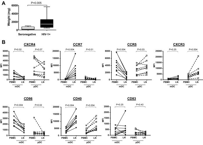
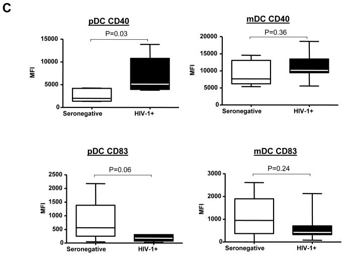
Dendritic cells (DCs) from HIV-1-infected individuals display numeric and functional defects, and recent evidence suggests that HIV-1 can directly and indirectly activate DCs in vitro. The in vivo activation state and compartmentalization of DC subsets during HIV-1 infection remain poorly understood, however. We evaluated phenotypic and functional characteristics of myeloid dendritic cells (mDCs) and plasmacytoid dendritic cells (pDCs) directly ex vivo in peripheral blood and lymphoid tissue from HIV-1-infected and HIV-seronegative individuals. Analysis of a wide range of chemokine receptors and activation/maturation markers on circulating DCs from viremic HIV-1-infected donors revealed a phenotype indicative of partial activation. Yet, blood DCs from viremic subjects still achieved full maturation when stimulated in vitro. In addition, blood pDCs from viremic individuals had a reduced capacity to migrate to CXCL12 in vitro. Total numbers of both DC subsets were increased in lymph nodes of asymptomatic untreated HIV-1-infected subjects, consistent with DC accumulation in the lymphoid compartment. Lymph node DCs also expressed high levels of CD40 in the absence of increases of other typical activation/maturation markers. Activation and depletion of DCs in blood with accumulation in lymphoid tissue may contribute to HIV-associated chronic immune activation and T-cell dysfunction.


Figures

FIGURE. 1
FIGURE. 1
Gating strategy to assess expression of various surface markers on DC subsets in blood and lymph nodes. Multi-parameter flow techniques were used to simultaneously assess expression of chemokine receptors (CCR5, CCR7, CXCR3, CXCR4) and costimulatory/activation markers (CD86, CD83, CD40) on both myeloid (mDC) and plasmacytoid (pDC) subsets. Total DCs, for both PBMC and lymph nodes, were defined as Lineage CD34 HLA-DR+ and then further divided into pDC (CD11c CD123high) and mDC (CD11chigh CD123low) subsets. Subsequent analysis of expression of chemokine and costimulatory/activation markers by these subsets was assessed. Net Mean Fluorescence Intensity (MFI) was determined using a fluorescence minus one (FMO) control for each fluorescent antibody. Profiles shown are representative of seronegative donors.
FIGURE 2
FIGURE 2
Expression of CD40 on myeloid (mDC) and plasmacytoid (pDC) DC subsets within freshly isolated peripheral blood and relationship to viral load. A) Expression of CD40 on mDC and pDC from seronegative (n=17), viremic (n=13) and suppressed (n=12) donors. Viral load is positively correlated with expression of B) CD40. Statistical analysis was performed using Kruskall-Wallis (KW) test for comparisons between multiple groups with pairwise comparisons conducted when P<0.05 using Dunn's multiple comparison adjustment and Spearman correlation test was performed for correlations. Values are expressed as Mean Fluorescence Intensity (MFI) minus FMO control as described in Fig. 1.
FIGURE 3
FIGURE 3
Migratory capacity of blood DCs from HIV-1-infected and seronegative subjects. A transwell migration assay was used to determine the ability of DC subsets to migrate to various chemokines. Values are displayed as net migration (percentage of DCs migrating to a chemokine minus the percent migrating to media alone). A) Comparisons of myeloid DC (mDC) and plasmacytoid DC (pDC) for seronegative donors (CXCL12 n=12; CCL5 n=10; CCL21 n=12; CXCL-10 n=10). B) Comparisons of mDC and pDC for viremic donors (CXCL12 n=16; CCL5 n=10; CCL21 n=15; CXCL-10 n=9). C) Net migration to CXCL12 for pDC from viremic donors compared to pDC from seronegative and suppressed donors. Statistical analysis was performed using Mann-Whitney for comparisons between mDC and pDC and Kruskall-Wallis (KW) test for comparisons between multiple groups with pairwise comparisons conducted when P<0.05 using Dunn's multiple comparison adjustment.
FIGURE 4
FIGURE 4
Comparison of lymph node weights and expression of surface markers on DC subsets from blood and lymph nodes from HIV-1-infected donors. A) Differences in overall weights (mg) of lymph nodes were compared between seronegative and HIV-1-infected (HIV-1+) subjects and statistical analysis performed using Mann-Whitney test. B) and C) Multi-parameter flow techniques were used to assess the expression of various chemokine receptors (CCR5, CCR7, CXCR3, CXCR4) and costimulatory/activation markers (CD86, CD83, CD40) on both myeloid DC (mDC) and plasmacytoid DC (pDC) subsets from freshly isolated peripheral blood (PBMC) and lymph nodes (LN) from 9 HIV-1-infected donors as shown in Fig.1. B) Changes in expression of markers by mDC and pDC within PBMC and lymph nodes (LN). Values are expressed as Mean Fluorescence Intensity (MFI) minus FMO control as described in Fig. 1. Statistical analysis was performed using Wilcoxon signed rank test. C) Expression of CD40 and CD83 on pDC and mDC in the lymph nodes of seronegative and HIV-1-infected (HIV-1+) donors. Values are expressed as Mean Fluorescence Intensity (MFI) minus FMO control (detailed in Fig. 1.). Statistical analysis was performed using Mann-Whitney test.
FIGURE 4
FIGURE 4
Comparison of lymph node weights and expression of surface markers on DC subsets from blood and lymph nodes from HIV-1-infected donors. A) Differences in overall weights (mg) of lymph nodes were compared between seronegative and HIV-1-infected (HIV-1+) subjects and statistical analysis performed using Mann-Whitney test. B) and C) Multi-parameter flow techniques were used to assess the expression of various chemokine receptors (CCR5, CCR7, CXCR3, CXCR4) and costimulatory/activation markers (CD86, CD83, CD40) on both myeloid DC (mDC) and plasmacytoid DC (pDC) subsets from freshly isolated peripheral blood (PBMC) and lymph nodes (LN) from 9 HIV-1-infected donors as shown in Fig.1. B) Changes in expression of markers by mDC and pDC within PBMC and lymph nodes (LN). Values are expressed as Mean Fluorescence Intensity (MFI) minus FMO control as described in Fig. 1. Statistical analysis was performed using Wilcoxon signed rank test. C) Expression of CD40 and CD83 on pDC and mDC in the lymph nodes of seronegative and HIV-1-infected (HIV-1+) donors. Values are expressed as Mean Fluorescence Intensity (MFI) minus FMO control (detailed in Fig. 1.). Statistical analysis was performed using Mann-Whitney test.

References

    1. Day CL, Kaufmann DE, Kiepiela P, et al. PD-1 expression on HIV-specific T cells is associated with T-cell exhaustion and disease progression. Nature. 2006 Sep 21;443(7109):350–354. - PubMed
    1. Meyaard L, Otto SA, Jonker RR, Mijnster MJ, Keet RP, Miedema F. Programmed death of T cells in HIV-1 infection. Science. 1992 Jul 10;257(5067):217–219. - PubMed
    1. Petrovas C, Casazza JP, Brenchley JM, et al. PD-1 is a regulator of virus-specific CD8+ T cell survival in HIV infection. J Exp Med. 2006 Oct 2;203(10):2281–2292. - PMC - PubMed
    1. Selliah N, Finkel TH. Biochemical mechanisms of HIV induced T cell apoptosis. Cell Death Differ. 2001 Feb;8(2):127–136. - PubMed
    1. Silvestri G, Fedanov A, Germon S, et al. Divergent host responses during primary simian immunodeficiency virus SIVsm infection of natural sooty mangabey and nonnatural rhesus macaque hosts. J Virol. 2005 Apr;79(7):4043–4054. - PMC - PubMed

Publication types