. 2008 May;118(5):821-31.
doi: 10.1097/MLG.0b013e318164d0aa.

Gene transfer in human vestibular epithelia and the prospects for inner ear gene therapy

Affiliations

Gene transfer in human vestibular epithelia and the prospects for inner ear gene therapy

Bradley W Kesser et al. Laryngoscope. 2008 May.

Abstract

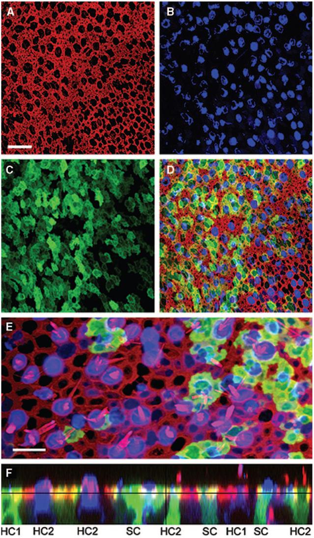
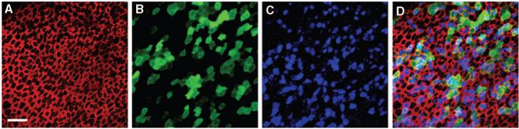
Transfer of exogenous genetic material into the mammalian inner ear using viral vectors has been characterized over the last decade. A number of different viral vectors have been shown to transfect the varying cell types of the nonprimate mammalian inner ear. Several routes of delivery have been identified for introduction of vectors into the inner ear while minimizing injury to existing structures and at the same time ensuring widespread distribution of the agent throughout the cochlea and the rest of the inner ear. These studies raise the possibility that gene transfer may be developed as a potential strategy for treating inner ear dysfunction in humans. Furthermore, a recent report showing successful transfection of excised human vestibular epithelia offers proof of principle that viral gene transfer is a viable strategy for introduction and expression of exogenous genetic material to restore function to the inner ear. Human vestibular epithelia were harvested from patients undergoing labyrinthectomy, either for intractable Ménière's disease or vestibular schwannoma resection, and cultured for as long as 5 days. In those experiments, recombinant, multiply-deleted, replication-deficient adenoviral vectors were used to transfect and express a reporter gene as well as the functionally relevant gene, wild-type KCNQ4, a potassium channel gene that when mutated causes the autosomal dominant HL DFNA2.Here, we review the current state of viral-mediated gene transfer in the inner ear and discuss different viral vectors, routes of delivery, and potential applications of gene therapy. Emphasis is placed on experiments demonstrating viral transfection of human inner ear tissue and implications of these findings and for the future of gene therapy in the human inner ear.


Figures

Fig. 1
Fig. 1
Confocal images of a human utricle harvested from patient 2. The explant was exposed to 107 viral particles/mL of a second generation, replication-deficient adenoviral vector containing the green fluorescent protein (GFP) gene insert for 15 hours and maintained in culture for 48 hours. The explant was then fixed and stained with Alexa Fluor 546-conjugated phalloidin and an antibody to myosin VIIa and an Alexa Fluor 633-conjugated secondary antibody. Scale bar = 20 μm and applies to A to D, which all show the same image field. (A) A stack of 12 images taken just beneath the apical surface of the epithelium was collapsed to show the cell bodies (B) and the subapical actin (red) that rings each hair cell and supporting cell. (B) Myosin VIIa immunoreactivity (blue) localizes sensory hair cell bodies. (C) Green fluorescence revealed that both hair cells and supporting cells were transfected by the vector and expressed GFP. (D) Merge of A to D. (E) Higher magnification overlay view from a different region of the same epithelium shown in panels A to D. To generate the image a stack of 13 images focused at the hair bundle level was collapsed (red=actin) and overlaid atop a collapsed stack of 10 images focused at the cell body level from the same image field (blue=myosin VIIa; green=GFP). Scale bar=10 μm. (F) A stack of 19 images projected to reveal a cross-sectional view of the sensory epithelium. Both type I (HC1) and type II hair cells (HC2) and supporting cells were GFP-positive. Myosin VIIa-positive hair cells are also evident. Same color code as for panels A to E. Reprinted from Kesser et al.
Fig. 2
Fig. 2
Confocal images of a saccule harvested from patient 1. The explant was cultured for 24 hours, fixed, and stained with Alexa Fluor 546 phalloidin (red) and endogenous KCNQ4 (green). (A) View from just above the apical surface of the epithelium reveals the actin-rich hair bundles. Scale bar = 25 μm and applies to A and B. (B) An image focused at the cell body level revealed the endogenous pattern of KCNQ4 expression. (C) Cross-sectional projection of the saccule generated from a stack of 43 images focused every 1.0 μm. The actin and KCNQ4 channels were merged. Scale bar = 10 μm. Reprinted from Kesser et al.
Fig. 3
Fig. 3
Stacks of confocal images taken just beneath the apical surface were used to generate images of the saccule harvested from a second patient undergoing labyrinthectomy for Ménière’s disease (patient 7). The explant culture was exposed to 2.3 × 10⁷ viral particles/mL of a second generation, replication-deficient adenovirus containing both the green fluorescent protein (GFP) and wild-type KCNQ4 gene inserts for 24 hours, fixed, and stained with Alex Fluor 546 phalloidin (red) and KCNQ4 (blue) and GFP (green). The scale bar represents 20 μm and applies to all images. The same field and focal planes are shown in A to D. (A) An image just beneath the apical surface of the epithelium shows the actin that rings each cell. (B) One hundred three GFP-positive cells are evident in this field. (C) The KCNQ4 antibody labeled both endogenous hair cell KCNQ4 and exogenous virally expressed KCNQ4. (D) Merge of A to C shows colocalization of GFP and KCNQ4 in 78.6% of the cells. Reprinted from Kesser et al.

References

    1. Lalwani AK, Castelein CM. Cracking the auditory genetic code: nonsyndromic hereditary hearing impairment. Am J Otol. 1999;20:115–132. - PubMed
    1. Raphael Y, Frisancho JC, Roessler BJ. Adenoviral-mediated gene transfer into guinea pig cochlear cells in vivo. Neurosci Lett. 1996;207:137–141. - PubMed
    1. Weiss MA, Frisancho JC, Roessler BJ, Raphael Y. Viral-mediated gene transfer in the cochlea. Int J Devel Neurosci. 1997;15:577–583. - PubMed
    1. Dazert Transfection of neonatal rat cochlear cells in vitro with an adenovirus vector. Int J Devel Neurosci. 1997;15:595–600. - PubMed
    1. Lalwani AK, Walsh BJ, Reilly PG, Muzyczka N, Mhatre AN. Development of in vivo gene therapy for hearing disorders: introduction of adeno-associated virus into the cochlea of the guinea pig. Gene Ther. 1996;3:588–592. - PubMed

MeSH terms

Substances