. 2008 Feb 27;3(2):e1653.
doi: 10.1371/journal.pone.0001653.

The Reelin receptors Apoer2 and Vldlr coordinate the patterning of Purkinje cell topography in the developing mouse cerebellum

Affiliations

The Reelin receptors Apoer2 and Vldlr coordinate the patterning of Purkinje cell topography in the developing mouse cerebellum

Matt Larouche et al. PLoS One. .

Abstract

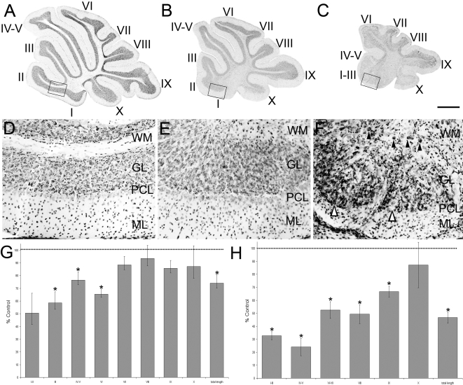
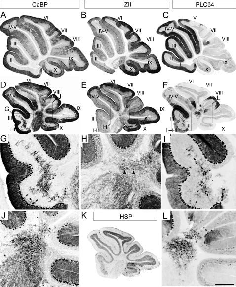
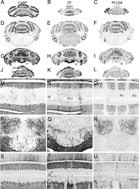
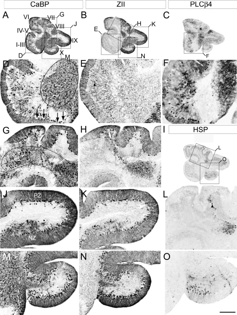
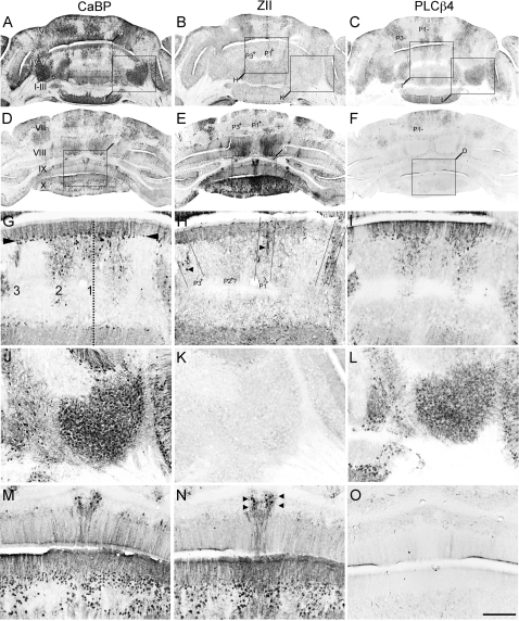
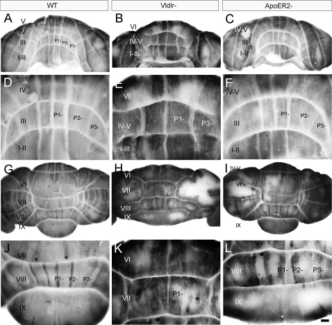
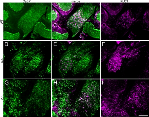
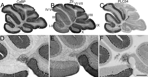
The adult cerebellar cortex is comprised of reproducible arrays of transverse zones and parasagittal stripes of Purkinje cells. Adult stripes are created through the perinatal rostrocaudal dispersion of embryonic Purkinje cell clusters, triggered by signaling through the Reelin pathway. Reelin is secreted by neurons in the external granular layer and deep cerebellar nuclei and binds to two high affinity extracellular receptors on Purkinje cells-the Very low density lipoprotein receptor (Vldlr) and apolipoprotein E receptor 2 (Apoer2). In mice null for either Reelin or double null for Vldlr and Apoer2, Purkinje cell clusters fail to disperse. Here we report that animals null for either Vldlr or Apoer2 individually, exhibit specific and parasagittally-restricted Purkinje cell ectopias. For example, in mice lacking Apoer2 function immunostaining reveals ectopic Purkinje cells that are largely restricted to the zebrin II-immunonegative population of the anterior vermis. In contrast, mice null for Vldlr have a much larger population of ectopic Purkinje cells that includes members from both the zebrin II-immunonegative and -immunopositive phenotypes. HSP25 immunoreactivity reveals that in Vldlr null animals a large portion of zebrin II-immunopositive ectopic cells are probably destined to become stripes in the central zone (lobules VI-VII). A small population of ectopic zebrin II-immunonegative Purkinje cells is also observed in animals heterozygous for both receptors (Apoer2(+/-): Vldlr(+/-)), but no ectopia is present in mice heterozygous for either receptor alone. These results indicate that Apoer2 and Vldlr coordinate the dispersal of distinct, but overlapping subsets of Purkinje cells in the developing cerebellum.


Conflict of interest statement

Competing Interests: The authors have declared that no competing interests exist.

Figures

Figure 1
Figure 1. Cresyl violet staining reveals that the cerebellar cortex of Apoer2 and Vldlr mutants is abnormal.
Sagittal sections through the medial cerebellum of adult wild type (A, D), Apoer2 (B, E) or Vldlr (C, F) null animals indicate that mutant cerebella are smaller and have fewer lobules when compared to wild type mice. Higher-power views reveal that a trilaminar structure is present in both mutants and wild type (D–F) consisting of an outer molecular layer (ML), Purkinje cell layer (PCL) and inner granule cell layer (GL). White matter tracts (WM) can also be observed in each animal. High magnification views of the Vldlr null cerebellum reveal the presence of Purkinje cell-sized somata in the granular layer and white matter (e.g. black arrowheads–F) as well as gaps in the Purkinje cell layer (white arrowheads–F). Measurements of the length of lobules in Apoer2 null (G) or Vldlr null (H) cerebella are expressed as a percentage of the length in wild-type littermates. Length measurements reveal a reduction in several areas of each mutant cerebellum. These reductions are most prominent in the anterior cerebellum of both mutants. Error bars on the graph depict SEM. Dotted line indicates the length of the equivalent lobule in wild type animals. Scale bar = 1 mm for A–C and 125 µm for D–F. * indicates p<0.05 as determined by one way ANOVA.
Figure 2
Figure 2. Adult Apoer2 null cerebella have Purkinje cell ectopia that is largely restricted to zebrin II-immunonegative cells.
Sagittal sections are taken from either adult wild type (A–C) or Apoer2 null (D–L) cerebella. Cerebella have been immunostained with antibodies against calbindin (CaBP-A, D, G, J), zebrin II (ZII-B, E, H), phospholipase Cß4 (PLCß4-C, F, I, L) or heat shock protein 25 (HSP25-K) to reveal immunopositive Purkinje cell bodies in the Purkinje cell layer (P) as well as their dendrites located within the molecular layer (M). Sections from the Apoer2 null cerebellum are serial sections (zebrin II-calbindin-PLCß4) while wild type sections are not. Boxes in D–F indicate areas where higher-magnification pictures are presented below. High-magnification panels (G, H, I, J, L) illustrate the presence of discrete groups of ectopic Purkinje cells in the white matter of the Apoer2 null cerebellum, as identified with CaBP-immunostaining (G, J).The absence of zebrin II immunoreactivity in these cells (H) indicates that the predominant phenotype of Purkinje cells in the white matter of these mutants is ZII-/PLC ß4+ (I, L). Black arrows in E point to areas in lobules IX and X where Purkinje cells are misaligned within the Purkinje cell monolayer. Arrowheads in H point to the rare occurrence of zebrin II immunopositive Purkinje cells in the ectopic clusters. K–HSP25 immunoreactivity is revealed in Purkinje cells throughout the NZ (dotted line). Roman numerals indicate lobules. Scale bar in L = 1 mm for A–F and 250 µm for E–L.
Figure 3
Figure 3. Purkinje cell ectopia is aligned with parasagittal organization in the aporER2 null cerebellum.
A series of transverse cryosections through adult wild type (A–C) or Apoer2 null (D–R) cerebella immunostained with antibodies against calbindin (CaBP-A, D, G, J, M, P), zebrin II (ZII-B, E, H, K, N, Q) or phospholipase Cß4 (PLCß4-C, F, I, L, O, R). Sections from the Apoer2 null cerebellum are serial (zebrin II-calbindin-PLCß4). Boxes in D–I indicate areas where higher-magnification photomicrographs are presented below as indicated by the label. High power views of anterior cerebella immunostained with CaBP (M) reveal that ectopic Purkinje cells are restricted to discrete parasagittal domains. Immunostaining with ZII and PLCß4 in neighboring sections reveals that these ectopic cells are composed of discrete and non-overlapping stripes of ectopic Purkinje cells (N, O). Ectopic Purkinje cells in the PZ (lobules VIII and dorsal IX) are from the zebrin II-negative subset (compare Q, R). Purkinje cell ectopia in the nodular zone (NZ) is limited to ZII positive Purkinje cells (S–U). Dotted line in panel T marks the approximate dorsoventral boundary within lobule IX between the posterior zone (including the dorsal half of lobule IX) and nodular zone (ventral IX and X). The transition between PZ and NZ in lobule IX highlights the differing characteristics of ectopic Purkinje cells in the NZ (misaligned ZII-positive) and PZ (ectopic clusters of ZII-negative Purkinje cells). P1+ and P2+ denote zebrin II-positive/PLCß4-negative Purkinje cell stripes, while P1- and P2- label zebrin II-negative/PLCß4-positive Purkinje cell stripes. Roman numerals indicate lobules. Scale bar in R = 1 mm for A–I and 250 µm for J–R.
Figure 4
Figure 4. Immunostaining of sagittal sections from adult Vldlr null cerebella reveals that Purkinje cell ectopia includes cells from both zebrin II-immunonegative and -immunopositive subsets.
A series of sagittal cryosections is illustrated from the vermis of adult Vldlr null cerebella immunostained for calbindin to reveal the location of all Purkinje cells (CaBP-A, D, G, J, M), as well as zebrin II (ZII-B, E, H, K, N), phospholipase C ß4 (PLCß4-C, F), or heat shock protein 25 (HSP25-I, L, O) to reveal the location of select subsets of Purkinje neurons. All four markers reveal that some Purkinje cells are correctly located within the Purkinje cell monolayer at the cerebellar cortex (e.g. between arrows–4D) as well as ectopically within the cerebellar white matter (e.g. 4D, G–dotted circles). The transition from posterior zone ( = PZ-lobule VIII and dorsal IX-Fig. 4J) into nodular zone ( = NZ-ventral IX and X–Fig. 4J) is revealed in the form of Purkinje cell ectopia (J, K). In the dorsal aspect of lobule IX Purkinje cells are restricted to a monolayer, with some ectopic cells located in the lobule white matter (J, K). In the ventral aspect of IX, the area of transition between the PZ->NZ, is highlighted by Purkinje cells misalignment and this misalignment extends the length of the NZ to include lobule X (Fig. 4M, N). Roman numerals denote putative lobule assignments. Scale bar = 1 mm for A–C and 250 µm for D–O.
Figure 5
Figure 5. Purkinje cell ectopia in the Vldlr null cerebellum is parasagittally organized.
Serial transverse cryosections through adult Vldlr null cerebellum immunostained with calbindin to reveal the location of all Purkinje cells (CaBP-A, D, G, J, M), or with zebrin II (ZII-B, E, H, K, N) phospholipase C ß4 (PLCß4-C, F, I, L, O) antibodies to reveal the location of parasagittal subsets of Purkinje cells. Boxes in A–F mark areas of higher magnification presented in the photomicrographs beneath as indicated by the letter on the corner of the box. Cells immunopositive for any of these three markers (CaBP, ZII, or PLCß4) are observed properly positioned within the Purkinje cell monolayer at the cerebellar cortex however numerous ectopic cells are also distributed throughout the cerebellar intralobular white matter. Most ectopic Purkinje cells in the anterior cerebellum are ZII-/PLCß4+ (5G–I). Some ZII-expressing Purkinje cells were observed in the granular layer (i.e. arrowheads–4E, 5H) and these ectopic Purkinje cells align in rough parasagittal stripes consistent with the overlying Purkinje cell topography in the cerebellar cortex (dotted lines-5H). M–O: high power views of ventral lobule IX (NZ) reveals that misaligned Purkinje cells are arranged into parasagittally-restricted groups, that are all zebrin II-positive (N). P1+, and P3+ mark zebrin II-immunopositive/PLCß4-immunonegative stripes and P1- and P2- mark zebrin II- immunonegative/PLCß4-immunopositive stripes. Arrowheads in G mark the Purkinje cell layer, the vertical dotted line denotes the midline, and numbers denote the location of ectopic clusters of Purkinje cells within the lobular white matter. Roman numerals denote cerebellar lobules. Scale bar in O = 1 mm for A–F and 250 µm for G–O.
Figure 6
Figure 6. PLCß4 immunostaining in Vldlr null whole mounts reveal patterning changes.
Whole cerebella from wild type (WT-A, D, G, J), Vldlr null (Vldlr-B, E, H, K) or Apoer2 null (ApoER2-C, F, I, L) cerebella immunostained with anti-PLCß4 antibodies. P1+, and P2+ mark zebrin II-immunopositive/PLCß4-immunonegative stripes while P1-, and P2- mark immunonegative/PLCß4-immunopositive stripes. The stripes are subtly altered in more dorsal lobules of the Vldlr- cerebellum. Specifically, as the P1- stripes enter putative lobule VI they narrow to ∼400 µm, while the P3- stripe remains roughly the same width as the AZ (∼600 µm) but veers sharply towards the hemispheres (E). PLCß4 whole mount immunostaining of the Apoer2 null cerebellum (C, F, I, L) reveals a parasagittal stripe pattern that is largely reminiscent of wild type and relatively unchanged despite the Purkinje cell ectopia observed inside the cerebellum (Fig 2, 3). Roman numerals (I–X) indicate lobules. Scale bar in L = 200 µm and applies to A–C, and G–I;  = 500 µm for G–F, and J–L.
Figure 7
Figure 7. KLC3 immunolabeling reveals that ectopic Purkinje cells do not intermingle with the deep cerebellar nuclei.
Sagittal cryosections from adult cerebella immunofluorescence labeled by using antibodies against KLC3 (magenta) to identify cerebellar nuclear neurons and calbindin (green) to identify Purkinje cells in wild type (A–C), Vldlr (D–F), or Apoer2 (G–I) null cerebella. Merged images reveal that ectopic Purkinje clusters lie near to, but outside of the deep cerebellar nuclei in both Apoer2 (A-/-) and Vldlr (V-/-) null mice. KLC-immunopositive cerebellar nuclear neurons are surrounded by the calbindin-immunoreactive axons of the Purkinje cells and these observations are consistent with previous studies of deep cerebellar nuclear neuron labeling (Chung et al., 2006). Wild type littermates (WT) have few ectopic Purkinje cells. Scale bar = 250 µm.
Figure 8
Figure 8. Cerebella from mice heterozygous for the Apoer2 and Vldlr deletions have Purkinje ectopia restricted to a small subset of zebrin II-immunonegative Purkinje cells.
Serial sagittal sections from adult cerebella immunoreacted with anti-calbindin (CaBP -A, E), anti-zebrin II (ZII-B, F), or anti-phospholipase C ß4 (PLCß4-C, G) antibodies reveal the presence of a small cluster of Purkinje cells (e.g. dotted circle–D) that fail to express zebrin II but do express PLCß4 (F). Roman numerals denote putative lobule assignments. Boxes in A–D indicate magnified areas. Scale bar in F = 1mm for A–C and 250 µm for D–F.

Similar articles

Cited by

References

    1. Hawkes R, Gravel C. The modular cerebellum. Prog Neurobiol. 1991;36:309–327. - PubMed
    1. Hawkes R. An anatomical model of cerebellar modules. Prog Brain Res. 1997;114:39–52. - PubMed
    1. Herrup K, Kuemerle B. The compartmentalization of the cerebellum. Ann Rev Neurosci. 1997;20:61–90. - PubMed
    1. Oberdick J, Baader SL, Schilling K. From zebra stripes to postal zones: deciphering patterns of gene expression in the cerebellum. Trends Neurosci. 1998;21:383–390. - PubMed
    1. Ozol K, Hayden J, Oberdick J, Hawkes R. Transverse zones in the vermis of the mouse cerebellum. J Comp Neurol. 1999;412:95–111. - PubMed

Publication types