. 2008 Jun 1;68(8):839-48.
doi: 10.1002/pros.20749.

Androgen receptor mediates the expression of UDP-glucuronosyltransferase 2 B15 and B17 genes

Affiliations

Androgen receptor mediates the expression of UDP-glucuronosyltransferase 2 B15 and B17 genes

Bo-Ying Bao et al. Prostate. .

Abstract

Background: Enhanced androgen receptor (AR) activity by increased testosterone availability may play important roles in prostate cancer progressing to castration resistant state. Comparison of expression profiles in androgen dependent and independent prostate tumors demonstrated a marked increase of the expression of UDP-glucuronosyltransferase 2B15 (UGT2B15), an androgen catabolic enzyme. We investigated mechanisms controlling the differential expression of UGT2B15 and B17 in response to androgen treatments.

Methods: Gene expression was determined by RT-PCR. The association of AR with UGT2B15/B17 genes was determined by Chromatin immuno-precipitation (CHIP). RNA interference was used to knock-down gene expression.

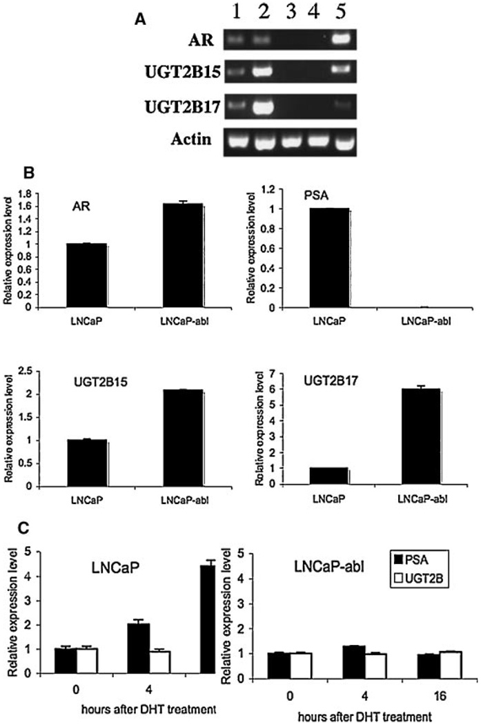
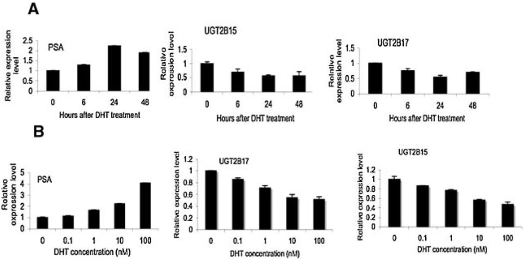
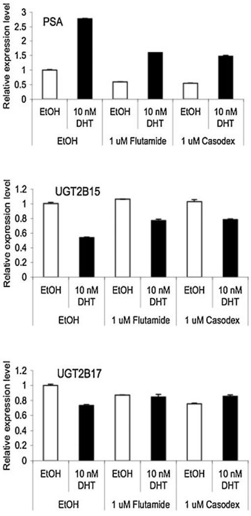
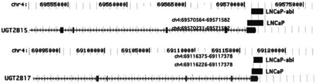
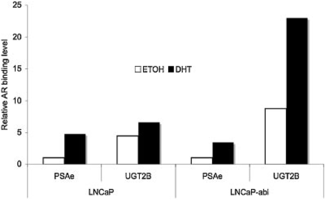
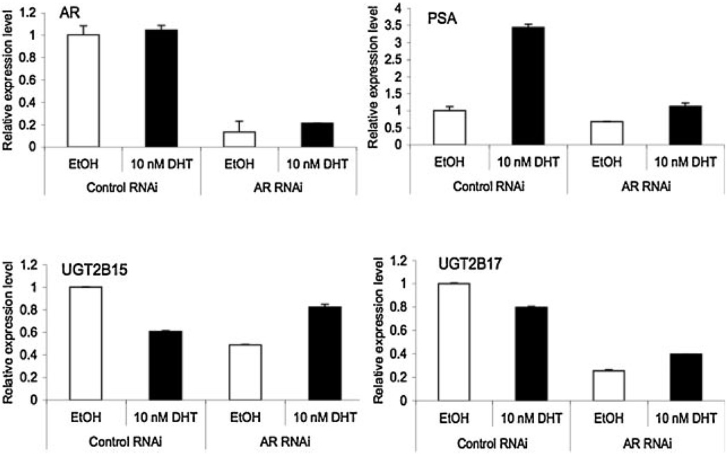
Results: UGT2B15 and B17 genes were not expressed in AR negative prostate cancer cell lines, PC3 and DU145, while they were expressed in AR positive cell lines, LNCaP, LNCaP-abl (an androgen independent LNCaP sub-line), and VCaP. The expression levels of UGT2B15/B17 were up-regulated in LNCaP-abl comparing to those in LNCaP. These results suggest the requirement of AR for the expression of UGT2B15/B17. Treatment with DHT down-regulated the expression of UGT2B15/B17 in LNCaP in a time and dose dependent manner and this down-regulation was competitively antagonized by flutamide and bicalutimide, suggesting a pathway mediated by AR. Further CHIP experiments demonstrated the direct interaction of AR with the promoter regions of UGT2B15/B17 genes. Knocking down AR expression in LNCaP significantly reduced the expression of UGT2B15/B17 and completely inhibited the DHT-induced down-regulation of UGT2B15/B17 genes.

Conclusions: We demonstrated that UGT2B15 and B17 are primary androgen-regulated genes and AR is required for both their basal expression and their androgen-regulated expression.


Figures

Fig. 1
Fig. 1
Differential expression of UGT2B15 and 17 in different PCa celllines. A: Comparison of UGT2B15 and 17 expression in AR positive and AR negative cell lines. Total RNAs were isolated from LNCaP (1), LNCaP-abl (2), DU145 (3),PC-3 (4), and VCaP (5) cell lines. Regular RT-PCR reactions were performed for ARUGT2B15, UGT2B17 and actin and the amplified cDNAs were separated on a 2.5% agarosegel. The expression level of actin was used as a control for estimating the relative amount of total RNAs applied in each reaction. B: Quantitative analysis of the expression level of UGT2B15 and B17 in LNCaP and LNCaP-abl cell lines. Real-time RT-PCR analysis was performed for measuring the relative mRNA level of AR, PSA, UGT2B15, and UGT2B17 in each cell line. Values represent the fold differences in gene expression relative to those in LNCaP cells, which was arbitrarily set as 1.0. C: Comparison of the response to DHT treatment in LNCaP and LNCaP-abl cell lines. Total RNAs were isolated from cells prior to the DHT treatment (0), and cells that were treated with 100 nMDHT for 4 hr (4) and 16 hr (16).The expression levels of PSA and UGT2B15/B17 were determined by real-time RT-PCR reactions. The level of expression in cells without DHT treatment (0) was arbitrarily set as 1.
Fig. 2
Fig. 2
Characterization of the impact of DHT treatment on UGT2B15 and B17 expression in LNCaP cells. A: Responses to different amounts of DHT. LNCaP cells were treated with different amounts of DHT, as indicated, for 24hr.B: Responses to the period of incubation time for DHT treatment. LNCaP cells were treated with 10 nMDHT for different periods of time, as indicated. Total RNAs were isolated and the expression levels of PSA, UGT2B15 and UGT2B17 were examined by real-time RT-PCR. Values represent the fold differences in gene expression relative to the expression level at 0 time point or without DHT treatment, which was arbitrarily set as 1.0.
Fig. 3
Fig. 3
The effect of anti-androgens on the DHT-induced down-regulation of UGT2B15/B17 expression. LNC aP cells were treated with ethanol vehicle,DHT (10nM), anti-androgens(Flutamide, 1µM; Casodex or bicalutamide, 1 µM), or a combination of DHT and anti-androgens for 24 hr and then total RNAs were isolated. The expression levels of PSA, UGT2B15, and UGT2B17 mRNAs were analyzed by real-time RT-PCR. Values represent the fold differences relative to those in cells without any drug treatments (which were set as 1.0).
Fig. 4
Fig. 4
Chromosomal locations of AR binding sites at the UGT2B15/B17 gene locus. UGT2B15 and B17 are two closely located genes at chromosome 4 (chr4). Numbers indicate the nucleotide positions on chr4. Physical maps of UGT2B15 and UGT2B17 genes are indicated by the arrowed-gray lines with gray vertical boxes, which represent exons and arrows indicate the transcription direction. Two black bars located on the top of the 5′ end of each gene represent the location of AR binding sites mapped in LNCaP and LNCaP-abl cell lines by ChIP-on-chip analysis.
Fig. 5
Fig. 5
CHIP analysis of the interaction of AR with the UGT2B15/B17 genelocus in LNCaP and LNCaP-abl celllines. Cells were treated with vehicle or 100 nMDHT for 16 hr, and then subjected to ChIP analyses using antibodies against AR following a previously established protocol [24,25].Immuno-precipitated DNAs derived from the UGT2B15/B17 locus were quantified by real-time PCR using primers spanning the 5′ end of the exon 1 of the UGT2B15/B17 genes, as described in Materials and Methods Section. The level of PSA enhancer (PSAe) bound with AR was simultaneously determined as a positive control, using the previously described primer set. In each experiment, the amount of input chromatin was measured by PCR amplification with the same set of primers and the amount of immuno-precipitated DNA was adjusted for the relative amount of input chromatin. Values represent the relative amount of AR bound material comparing to the level of AR bound PSAe in the absence of DHT, which was arbitrarily set as 1. Each value represents the mean from two independent experiments.
Fig. 6
Fig. 6
The impact of ARRNAion UGT2B15 and B17 expression. LNCaP cells were transfected with control and ARRNAi, respectively. Forty eight hours after transfection, cells were treated with ethanol vehicle or 10 nM DHT for another 24hr, and then total RNAs were isolated. AR, PSA, UGT2B15, and UGT2B17 mRNA expression were analyzed by real-time RT-PCR. Values represent the fold differences in gene expression relative to control. Each value represents the mean from two independent experiments.

Similar articles

Cited by

References

    1. Heinlein CA, Chang C. Androgen receptor in prostate cancer. Endocr Rev. 2004;25(2):276–308. - PubMed
    1. Gelmann EP. Molecular biology of the androgen receptor. J Clin Oncol. 2002;20:3001–3015. - PubMed
    1. Feldman BJ, Feldman D. The development of androgen-independent prostate cancer. Nat Rev Cancer. 2001;1(1):34–45. - PubMed
    1. Balk SP. Androgen receptor as a target in androgen independent prostate cancer. Urology. 2002;60:132–138. - PubMed
    1. Holzbeierlein J, Lal P, LaTulippe E, Smith A, Satagopan J, Zhang L, Ryan C, Smith S, Scher H, Scardino P, Reuter V, Gerald WL. Gene expression analysis of human prostate carcinoma during hormonal therapy identifies androgen-responsive genes and mechanisms of therapy resistance. Am J Pathol. 2004;164:217–227. - PMC - PubMed

Publication types

MeSH terms

Substances