. 2008 Feb 27:5:32.
doi: 10.1186/1743-422X-5-32.

Mapping of immunogenic and protein-interacting regions at the surface of the seven-bladed beta-propeller domain of the HIV-1 cellular interactor EED

Affiliations

Mapping of immunogenic and protein-interacting regions at the surface of the seven-bladed beta-propeller domain of the HIV-1 cellular interactor EED

Dina Rakotobe et al. Virol J. .

Abstract

Background: The human EED protein, a member of the superfamily of Polycomb group proteins, is involved in multiple cellular protein complexes. Its C-terminal domain, which is common to the four EED isoforms, contains seven repeats of a canonical WD-40 motif. EED is an interactor of three HIV-1 proteins, matrix (MA), integrase (IN) and Nef. An antiviral activity has been found to be associated with isoforms EED3 and EED4 at the late stage of HIV-1 replication, due to a negative effect on virus assembly and genomic RNA packaging. The aim of the present study was to determine the regions of the EED C-terminal core domain which were accessible and available to protein interactions, using three-dimensional (3D) protein homology modelling with a WD-40 protein of known structure, and epitope mapping of anti-EED antibodies.

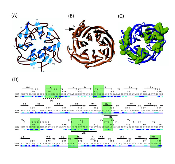
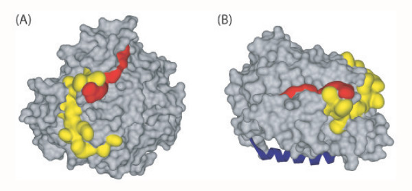
Results: Our data suggested that the C-terminal domain of EED was folded as a seven-bladed beta-propeller protein. During the completion of our work, crystallographic data of EED became available from co-crystals of the EED C-terminal core with the N-terminal domain of its cellular partner EZH2. Our 3D-model was in good congruence with the refined structural model determined from crystallographic data, except for a unique alpha-helix in the fourth beta-blade. More importantly, the position of flexible loops and accessible beta-strands on the beta-propeller was consistent with our mapping of immunogenic epitopes and sites of interaction with HIV-1 MA and IN. Certain immunoreactive regions were found to overlap with the EZH2, MA and IN binding sites, confirming their accessibility and reactivity at the surface of EED. Crystal structure of EED showed that the two discrete regions of interaction with MA and IN did not overlap with each other, nor with the EZH2 binding pocket, but were contiguous, and formed a continuous binding groove running along the lateral face of the beta-propeller.

Conclusion: Identification of antibody-, MA-, IN- and EZH2-binding sites at the surface of the EED isoform 3 provided a global picture of the immunogenic and protein-protein interacting regions in the EED C-terminal domain, organized as a seven-bladed beta-propeller protein. Mapping of the HIV-1 MA and IN binding sites on the 3D-model of EED core predicted that EED-bound MA and IN ligands would be in close vicinity at the surface of the beta-propeller, and that the occurrence of a ternary complex MA-EED-IN would be possible.


Figures

Figure 1
Figure 1
Crystallogenesis of histidine-tagged isoform 3 of EED. (A), Platelet-like crystals of EED3-H6 protein of 441 residues, obtained in suspended drop in 0.1 M MES buffer pH 6.0, 40 % MPD. (B), Solubilization of the crystals and analysis by SDS-PAGE and Coomassie blue staining. Lane 1 : solution of purified EED3-H6 (10 mg/mL) used for crystallogenesis (protein load : 50 μg). Lane 2 : protein content of solubilized single crystal. MW : markers of protein molecular mass, indicated in kilodaltons (kDa) on the right side of the panel.
Figure 2
Figure 2
Structural models and immunogenic regions of EED isoform 3. (A), Seven-bladed β-propeller model of the EED core domain, based on sequence homology with the beta subunit of the bovine G protein (Gβ ; [27, 28]). Shown is a ribbon representation of the polypeptide backbone atoms of EED3 isoform (amino acid residues 84–441), with secondary and tertiary structures of the different β-blades. (B), 3D-model of the EED3 seven-bladed β-propeller, deduced from crystallographic data (modified, from [30]). The black arrow indicates the major difference between our putative model (A) and the crystal model (B), consisting of the α1 helical region facing the β-strand β17 in β-blade IV. (C), Position of immunogenic epitopes (depicted in green) on the 3D-model of EED polypeptide backbone (represented in blue). (D), Primary and secondary structures of EED3, deduced from crystallographic data [30]. The amino acid sequence was numbered according to the accepted nomenclature [12] : Met95 in EED1 isoform represented Met1 in EED3 ; thus, the C-terminal residue L440 in EED3 corresponded to L535 in EED1. Regions in β-strand structure are represented by horizontal arrows, with reference to the blade number and β-strand letter a, b, c or d ; α-helices are represented by spirals, and turns by TT. Helical regions marked α1 and η1, and the β-strand region marked β17, were structurized domains of EED which were unique among representatives of WD-40 proteins. The relative accessibility of each residue (acc) in the 3D structure was extracted from the dictionary of protein structure [45], and indicated as coloured bars under the sequence with the following colour code : dark blue, highly accessible ; light blue, accessible ; white, buried. Discrete regions recognized by anti-EED IgG are indicated by green boxes. The binding sites of HIV-1 matrix protein (MA) and integrase (IN) are underlined by solid black lines.
Figure 3
Figure 3
Surface representation of the β-propeller domain of EED and protein-interacting regions. The binding residues of HIV-1 proteins are represented with the following colour code : yellow for the matrix protein (MA), red for integrase (IN). (A), Top view of the β-propeller showing the MA and IN binding sites laterally oriented. Note the absence of overlapping between the MA and IN binding sites, which form a continuous binding groove. (B), Side view of the β-propeller showing the MA+IN-binding groove on the lateral face, and the position of the EZH2 α-helical peptide 39–68 (represented in blue), bound to the EZH2-binding pocket facing downwards.

Similar articles

Cited by

References

    1. Ng J, Li R, Morgan K, Simon J. Evolutionary conservation and predicted structure of the Drosophila extra sex combs repressor protein. Mol Cell Biol. 1997;17:6663–6672. - PMC - PubMed
    1. Schumacher A, Faust C, Magnuson T. Positional cloning of a global regulator of anterior-posterior patterning in mice. Nature. 1996;383:250–253. doi: 10.1038/383250a0. - DOI - PubMed
    1. Schumacher A, Lichtarge O, Schwartz S, Magnusson T. The murine polycombgroup eed and its human orthologue: functional implications of evolutionary conservation. Genomics. 1998;54:79–88. doi: 10.1006/geno.1998.5509. - DOI - PubMed
    1. Simon J. Locking in stable states of gene expression: transcriptional control during Drosophila development. Curr Opin Cell Biol. 1995;7:376–385. doi: 10.1016/0955-0674(95)80093-X. - DOI - PubMed
    1. Sewalt RGAB, van der Vlag J, Gunster MJ, Hamer KM, den Blaauwen JL, Satijn DPE, Hendrix T, van Driel R, Otte AP. Characterization of interactions between the mammalian Polycomb-group proteins Enx1/EZH2 and EED suggests the existence of different mammalian Polycomb-group protein complexes. Mol Cell Biol. 1998;18:3586–3595. - PMC - PubMed

Publication types

MeSH terms

LinkOut - more resources