Review
. 2008;18(2):139-50.
doi: 10.1615/critreveukargeneexpr.v18.i2.30.

Regulation of chondrocytic gene expression by biomechanical signals

Affiliations
Review

Regulation of chondrocytic gene expression by biomechanical signals

Thomas J Knobloch et al. Crit Rev Eukaryot Gene Expr. 2008.

Abstract

Cartilage is a mechanosensitive tissue, which means that it can perceive and respond to biomechanical signals. Despite the known importance of biomechanical signals in the etiopathogenesis of arthritic diseases and their effectiveness in joint restoration, little is understood about their actions at the cellular level. Recent molecular approaches have revealed that specific biomechanical stimuli and cell interactions generate intracellular signals that are powerful inducers or suppressors of proinflammatory and reparative genes in chondrocytes. Biomechanical signals are perceived by cartilage in magnitude-, frequency-, and time-dependent manners. Static and dynamic biomechanical forces of high magnitudes induce proinflammatory genes and inhibit matrix synthesis. Contrarily, dynamic biomechanical signals of low/physiologic magnitudes are potent antiinflammatory signals that inhibit interleukin-1beta (IL-1beta)-induced proinflammatory gene transcription and abrogate IL-1beta/tumor necrosis factor-alpha-induced inhibition of matrix synthesis. Recent studies have identified nuclear factor-kB (NF-kB) transcription factors as key regulators of biomechanical signal-mediated proinflammatory and antiinflammatory actions. These signals intercept multiple steps in the NF-kappaB signaling cascade to regulate cytokine gene expression. Taken together, these findings provide insight into how biomechanical signals regulate inflammatory and reparative gene transcription, underscoring their potential in enhancing the ability of chondrocytes to curb inflammation in diseased joints.


Figures

FIGURE 1
FIGURE 1
Schematic representation of types of biomechanical forces exerted on chondrocytes. (A) A normal chondrocyte in a lacunae surrounded by extracellular matrix (ECM). (B) Deformation of chondrocytes and ECM during active compression that consequently resulted in passive tensile forces, radial fluid flow, and increased nutrient transport. (C) Deformation of chondrocytes and ECM in response to shear forces that resulted in minimal hydrostatic pressure, fluid flow, and nutrient transport. (D) Schematic representation of dynamic compressive forces that can be of equal time intervals or of varying time intervals (frequencies). Dynamic compression leads to cyclic matrix and chondrocyte deformation, changes in hydrostatic pressure, and enhanced flow of nutrients. (E) Static compression showing ramp- and hold-type effects that increase hydrostatic pressure and induce matrix deformation that is followed by static conditions and minimal transport of nutrients.,
FIGURE 2
FIGURE 2
Transcriptional regulation of proinflammatory and reparative genes in response to various types of mechanical forces. Dynamic compressive and tensile forces of high magnitudes and static forces induce expression of proinflammatory genes that are associated with matrix destruction such as inducible nitric oxide synthase, cyclooxygenase 2, matrix metalloproteinases, a disintegrin-like and metalloprotease domain (reprolysin-type) with thrombospondin type I motifs 4 (ADAMTS4), and ADAMTS5. In parallel, these forces inhibit expression of matrix-associated molecules such as aggrecan, collagen type II (COL2), tissue inhibitor of metalloproteinases (TIMPs), and cartilage oligomeric matrix protein (COMP). On the other hand, dynamic compressive and tensile forces of low magnitudes upregulate synthesis of matrix-associated proteins such as proteoglycans, COL2, aggrecan, TIMPs, and COMP. In addition, biomechanical signals of low magnitudes inhibit interleukin-1β–induced expression of proinflammatory genes as well as abrogate cytokine-mediated inhibition of synthesis of matrix-associated proteins.
FIGURE 3
FIGURE 3
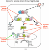
Schematic representation of the mechanisms of intracellular actions of dynamic tensile strain (DTS). (A) DTS of low magnitudes (DTS-L) suppresses interleukin-1β (IL-1β)–induced proinflammatory gene induction by intercepting salient steps in the nuclear factor-κB (NF-κB) signaling cascade to inhibit its transcriptional activity. (1) DTS suppresses IL-1β–induced inhibitor of κ light polypeptide gene enhancer in B cells kinase activation and thus phosphorylation and proteosomal degradation of I-κBα and I-κBβ. This leads to the inhibition of NF-κB nuclear translocation. (2) During the initial stages of IL-1β–mediated activation of cells, DTS upregulates I-κBβ nuclear translocation to prevent NF-κB binding to the DNA and to facilitate export of nuclear NF-κB, which may enter the nucleus. (3) DTS represses IL-1β–induced I-κBα and I-κBβ mRNA expression. Collectively, these actions of DTS inhibit proinflammatory gene induction as well as expression of multiple molecules involved in regulation of the NF-κB signaling cascade to suppress IL-1β–induced inflammation. (B) DTS of high magnitudes (DTS-H) upregulates proinflammatory gene transcription by inducing I-κBα and I-κBβ degradation and subsequent nuclear translocation of NF-κB. This results in the transcriptional activation of proinflammatory mediators including nitric oxide synthase 2, cyclooxygenase 2, matrix metalloproteinases, IL-1β, and tumor necrosis factor-α (TNF-α) and inhibition of the expression of matrix-associated proteins aggrecan, collagen type II, and tissue inhibitor of metalloproteinases. Black arrows indicate IL-1β–mediated activation of NF-κB transcription factors. Red arrows indicate steps in the NF-κB cascade that are inhibited by DTS-L, and the green arrows indicate points that are upregulated by DTS-L.
FIGURE 3
FIGURE 3
Schematic representation of the mechanisms of intracellular actions of dynamic tensile strain (DTS). (A) DTS of low magnitudes (DTS-L) suppresses interleukin-1β (IL-1β)–induced proinflammatory gene induction by intercepting salient steps in the nuclear factor-κB (NF-κB) signaling cascade to inhibit its transcriptional activity. (1) DTS suppresses IL-1β–induced inhibitor of κ light polypeptide gene enhancer in B cells kinase activation and thus phosphorylation and proteosomal degradation of I-κBα and I-κBβ. This leads to the inhibition of NF-κB nuclear translocation. (2) During the initial stages of IL-1β–mediated activation of cells, DTS upregulates I-κBβ nuclear translocation to prevent NF-κB binding to the DNA and to facilitate export of nuclear NF-κB, which may enter the nucleus. (3) DTS represses IL-1β–induced I-κBα and I-κBβ mRNA expression. Collectively, these actions of DTS inhibit proinflammatory gene induction as well as expression of multiple molecules involved in regulation of the NF-κB signaling cascade to suppress IL-1β–induced inflammation. (B) DTS of high magnitudes (DTS-H) upregulates proinflammatory gene transcription by inducing I-κBα and I-κBβ degradation and subsequent nuclear translocation of NF-κB. This results in the transcriptional activation of proinflammatory mediators including nitric oxide synthase 2, cyclooxygenase 2, matrix metalloproteinases, IL-1β, and tumor necrosis factor-α (TNF-α) and inhibition of the expression of matrix-associated proteins aggrecan, collagen type II, and tissue inhibitor of metalloproteinases. Black arrows indicate IL-1β–mediated activation of NF-κB transcription factors. Red arrows indicate steps in the NF-κB cascade that are inhibited by DTS-L, and the green arrows indicate points that are upregulated by DTS-L.

Similar articles

Cited by

References

    1. Grodzinsky AJ, Levenston ME, Jin M, Frank EH. Cartilage tissue remodeling in response to mechanical forces. Annu Rev Biomed Eng. 2000;2:691–713. - PubMed
    1. Park S, Hung CT, Ateshian GA. Mechanical response of bovine articular cartilage under dynamic unconfined compression loading at physiological stress levels. Osteoarthritis Cartilage. 2004;12(1):65–73. - PubMed
    1. Fehrenbacher A, Steck E, Rickert M, Roth W, Richter W. Rapid regulation of collagen but not metalloproteinase 1, 3, 13, 14 and tissue inhibitor of metalloproteinase 1, 2, 3 expression in response to mechanical loading of cartilage explants in vitro. Arch Biochem Biophys. 2003;410(1):39–47. - PubMed
    1. Fitzgerald JB, Jin M, Grodzinsky AJ. Shear and compression differentially regulate clusters of functionally related temporal transcription patterns in cartilage tissue. J Biol Chem. 2006;281(34):24095–103. - PubMed
    1. Giannoni P, Siegrist M, Hunziker EB, Wong M. The mechanosensitivity of cartilage oligomeric matrix protein (COMP) Biorheology. 2003;40(1–3):101–9. - PubMed

Publication types