. 2008 Mar;212(3):211-28.
doi: 10.1111/j.1469-7580.2008.00864.x.

Structure-function relationships in tendons: a review

Affiliations

Structure-function relationships in tendons: a review

M Benjamin et al. J Anat. 2008 Mar.

Abstract

The purpose of the current review is to highlight the structure-function relationship of tendons and related structures to provide an overview for readers whose interest in tendons needs to be underpinned by anatomy. Because of the availability of several recent reviews on tendon development and entheses, the focus of the current work is primarily directed towards what can best be described as the 'tendon proper' or the 'mid-substance' of tendons. The review covers all levels of tendon structure from the molecular to the gross and deals both with the extracellular matrix and with tendon cells. The latter are often called 'tenocytes' and are increasingly recognized as a defined cell population that is functionally and phenotypically distinct from other fibroblast-like cells. This is illustrated by their response to different types of mechanical stress. However, it is not only tendon cells, but tendons as a whole that exhibit distinct structure-function relationships geared to the changing mechanical stresses to which they are subject. This aspect of tendon biology is considered in some detail. Attention is briefly directed to the blood and nerve supply of tendons, for this is an important issue that relates to the intrinsic healing capacity of tendons. Structures closely related to tendons (joint capsules, tendon sheaths, pulleys, retinacula, fat pads and bursae) are also covered and the concept of a 'supertendon' is introduced to describe a collection of tendons in which the function of the whole complex exceeds that of its individual members. Finally, attention is drawn to the important relationship between tendons and fascia, highlighted by Wood Jones in his concept of an 'ectoskeleton' over half a century ago - work that is often forgotten today.


Figures

Fig. 1
Fig. 1
A sagittal section through the knee joint showing the presence of intramuscular tendons (arrows) within the muscle belly of the gastrocnemius (G) and hamstring (H) muscles. F, femur; QT, quadriceps tendon; P, patella; PT, patellar tendon; T, tibia.
Fig. 2
Fig. 2
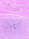
(a) A low power, longitudinal section through the limb tendon of a young calf in a section stained with Haematoxylin and Eosin (H & E). The tenocytes (TC) are typically arranged in longitudinal rows between parallel bundles of collagen fibres (CF) and are only recognizable in such routine sections by their darkly staining nuclei (i.e. the cytoplasm is not visible). Note the waviness (crimp) of the collagen. (b) A low power transverse section through the limb tendon of a young calf stained with H & E. Note that the collagen fibres are grouped into fascicles (FA) separated by endotenon (E). The tenocytes are recognizable within the fascicles by their nuclei (arrows).
Fig. 3
Fig. 3
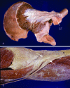
(a) The aponeurotic tendon (arrows) of gluteus minimus emerging from the surface of the muscle and attaching to the greater trochanter (GT) of the femur. I, ilium. (b) The pes anserinus tendon complex attaching to the tibia (T). Note the aponeurotic character of the distal part of the tendons (arrows). G, gastrocnemius; S, sartorius.
Fig. 4
Fig. 4
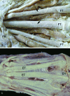
The gross anatomy of tendons in the hand. (a) The flexor digitorum superficialis tendons (FT) emerging from beneath the flexor retinaculum (FR) to enter the palm of the hand. Note their rounded character and the shallow grooves that are occasionally evident on their surface (arrows). L, lumbricals. (b) The web of extensor tendons (ET) on the dorsum of the hand collectively form a ‘supertendon’ complex in which the individual components are interconnected by films of connective tissue (CT) and obliquely-orientated juncturae tendinum (JT). Note the extensor hood (EH) over the metacarpophalangeal joints.
Fig. 5
Fig. 5
(a) A sagittal section of the attachment of the Achilles tendon (AT) to the calcaneus (C), showing the relation of it to the superior tuberosity (ST) that acts as a tendon pulley during dorsiflexion. Note also the presence of Kager's fat pad (KP) filling the space between the Achilles tendon and flexor hallucis longus (FHL). It contains numerous blood vessels (arrows), some of which enter the deep surface of the Achilles tendon to supply it. (b) A sagittal section of a toe that is hyperextended at the metatarsophalangeal joint (MTJ) and flexed at both interphalangeal joints (IPJ). Note how the head of the metatarsal acts as a pulley not only for the plantar fascia in maintaining the medial longitudinal arch of the foot, but also for the flexor tendons (FT) when the phalanges are dorsiflexed at the MTJ.
Fig. 6
Fig. 6
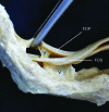
Vinculae (V) associated with the tendons of flexor digitorum superficialis (FDS) and flexor digitorum profundus (FDP) in a finger. The vinculae are remnants of the mesotenon and convey blood vessels to the tendons.
Fig. 7
Fig. 7
A sagittal section through the interphalangeal joint of the thumb stained with Masson's trichrome, showing how the tendon of extensor pollicis longus (EPL) replaces the joint capsule dorsally. DP, distal phalanx; PP, proximal phalanx.
Fig. 8
Fig. 8
The biceps brachii muscle (BM) of the forearm has a tendon that attaches to the bicipital tuberosity of the radius (R) and an aponeurotic expansion (A) that merges with the deep fascia of the forearm. The bicipital bursa (BB) has been opened up at the tendon attachment site.
Fig. 9
Fig. 9
Two examples of tendons that have completely abandoned a bony enthesis and are attached to fascia instead. (a) Tensor fascia latae (TFL) attaching to the iliotibial tract (ITT). (b) The tendon of palmaris longus (PL) attaching to the palmar aponeurosis (PA).
Fig. 10
Fig. 10
(a) A lateral view of the dorsal digital expansion (DE) of a finger on the proximal phalanx. Note the presence of interosseous (I) and lumbrical (L) muscles that attach to the expansion and the existence of a fibrous flexor sheath (FS) on the palmar aspect of the finger. (b) A dorsal view of the dorsal digital expansion over a metacarpophalangeal joint and its relationship to the more proximal extensor tendon (ET). The lateral slips of the extensor expansion have been displayed over the intermediate phalanx (arrows). (c) The three ‘wrap-around tendons’ in the region of the medial malleolus (MM) – tibialis posterior (TP), flexor digitorum longus (FDL) and flexor hallucis longus (FHL). The tendon of tibialis posterior has been displaced from its groove (arrow) to show the ‘articular character’ of the bone surface against which it presses. The left side of the photograph is distal. (d) The quadratus plantae muscle (QP) in the sole of the foot. It serves to adjust the oblique pull of flexor digitorum longus (FDL).
Fig. 11
Fig. 11
A histological view of a region of a tendon subject to compression (extensor pollicis longus as it crosses the interphalangeal joint of the thumb). Fibrocartilage cells (FC) rather than fibroblasts are typical of the compressed region of the tendon. Toluidine blue stained section.
Fig. 12
Fig. 12
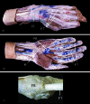
(a,b) Synovial sheaths associated with tendons in the wrist and hand as demonstrated in specimens injected with a blue dye. Note the presence of such sheaths around the digital flexor tendons (FT), but their absence in association with the digital extensor tendons (ET). On both the extensor (a) and flexor (b) sides, the synovial sheaths extend beyond the limits of the wrist retinacula (R) to allow for the longitudinal excursion of the tendons. The palmar bursa (PB) is evident in (b). (c) The digital flexor tendons (FT) entering the tunnel created by the fibrous flexor sheath (FS) and the phalangeal bones in a finger.

Similar articles

Cited by

References

    1. Ackermann PW, Li J, Finn A, Ahmed M, Kreicbergs A. Autonomic innervation of tendons, ligaments and joint capsules. A morphologic and quantitative study in the rat. J Orthop Res. 2001;19:372–378. - PubMed
    1. Alexander RM. Elastic mechanisms in primate locomotion. Z Morphol Anthropol. 1991;78:315–320. - PubMed
    1. Alfredson H, Lorentzon R. Sclerosing polidocanol injections of small vessels to treat the chronic painful tendon. Cardiovasc Hematol Agents Med Chem. 2007;5:97–100. - PubMed
    1. Alm A, Stromberg B. Vascular anatomy of the patellar and cruciate ligaments. A microangiographic and histologic investigation in the dog. Acta Chir Scand Suppl. 1974;445:25–35. - PubMed
    1. Amadio PC. Friction of the gliding surface. Implications for tendon surgery and rehabilitation. J Hand Ther. 2005;18:112–119. - PMC - PubMed

LinkOut - more resources