. 2008 Mar;212(3):257-74.
doi: 10.1111/j.1469-7580.2008.00861.x.

Unique features of spermiogenesis in the Musky Rat-kangaroo: reflection of a basal lineage or a distinct fertilization process?

Affiliations

Unique features of spermiogenesis in the Musky Rat-kangaroo: reflection of a basal lineage or a distinct fertilization process?

Shan Lloyd et al. J Anat. 2008 Mar.

Abstract

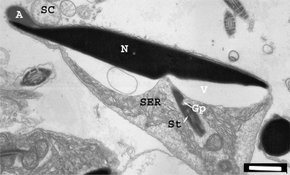
Previous research has found the mature spermatozoon of the Musky Rat-kangaroo to share many characteristics with other macopodoids, some phalangeroids and even the dasyurids. While there have been several studies published on the ultrastructure of the mature marsupial spermatozoon, there are only a few detailed studies on marsupial spermatogenesis. Furthermore, there have been no studies undertaken which combine the staging of the epithelial cell cycle with transmission electron microscopy to describe the ultrastructural changes in the developing spermatozoon during these stages. Such studies have the potential to be used in determining the required time taken for certain components of the spermatozoa to develop. During this study, eight stages of the seminiferous epithelium were observed and the ultrastructure of spermatogenesis was divided into nine phases. Maturational processes in the epididymides are also described. Among the features reported are: the formation of a unique acrosomal granule different from those reported in any other marsupial, the absence of contraction of the nuclear ring, a conspicuous acrosomal compaction process despite the almost 100% coverage of the dorsal nuclear surface and the retention of late spermatids within the seminiferous tubules until the early spermatids have developed to the nuclear protrusion phase.


Figures

Fig. 1
Fig. 1
Pro-acrosomal vacuole. (a) Section through the early spermatid just after spermatocyte division. Early flagellum arrowed. Inset: intercellular bridge between spermatids. (b) Spermatid in a slightly later stage of development than in (a) showing an active Golgi. A, acrosome; AG, acrosomal granule; An, annulus; B, intercellular bridge; G, Golgi apparatus; Gr, granular material; L, lateral junctional body; LC, longitudinal centriole; N, nucleus; NM, nuclear membrane; PaV, pro-acrosomal vacuole; PJB, proximal junctional body; TC, transverse centriole. Scale bars: (a,b) 1 µm; (a, inset) 500 nm.
Fig. 9
Fig. 9
Spermiation. The tail has rotated again to leave the neck and head at an angle of about 45°. The spermatid is released from the Sertoli cell cytoplasm, a small droplet remaining above the acrosome. DP, distal portion of the proximal junctional body; Gp, gap; Mi, mitochondria; PP, proximal portion of the proximal junctional body; SC, Sertoli cell cytoplasm; SER, smooth endoplasmic reticulum; St, striated sheath. For other abbreviations see Figs 1–6. Scale bar: 1 µm.
Fig. 2
Fig. 2
Acrosomal vacuole. (a) Shows a spermatid at the beginning of this phase. Several small vesicles from Golgi have amalgamated to form the acrosomal vacuole. (b) The vacuole begins to indent the nucleus and a granular material is deposited on the acrosomal membrane adjacent to the nucleus. (c–g) As the acrosome enlarges further, a membranous granule, which takes on a variety of forms as illustrated in these micrographs, is evident. (c, inset) Developing neck structures during this phase. See Fig. 1 for abbreviations. Scale bars: (a,c,f), 500 nm; (b,d,e,g), 1 µm, (c, inset), 100 nm.
Fig. 2
Fig. 2
Acrosomal vacuole. (a) Shows a spermatid at the beginning of this phase. Several small vesicles from Golgi have amalgamated to form the acrosomal vacuole. (b) The vacuole begins to indent the nucleus and a granular material is deposited on the acrosomal membrane adjacent to the nucleus. (c–g) As the acrosome enlarges further, a membranous granule, which takes on a variety of forms as illustrated in these micrographs, is evident. (c, inset) Developing neck structures during this phase. See Fig. 1 for abbreviations. Scale bars: (a,c,f), 500 nm; (b,d,e,g), 1 µm, (c, inset), 100 nm.
Fig. 3
Fig. 3
Nuclear protrusion. The acrosome has collapsed and the spermatid has taken on a triangular appearance with the nucleus forming the apex. The nuclear membrane is thickened beneath the acrosome, marked by the white arrow and the nuclear membrane is turned in at the acrosomal edge (black arrow). (Inset) This micrograph clearly demonstrates the nuclear, acrosome, plasma and Sertoli cell membranes as well as the electron-dense narrow band (white arrow) between the nuclear and acrosomal membranes. Scale bars: 1 µm, (inset) 100 nm.
Fig. 4
Fig. 4
Nuclear flattening. (a) The nucleus has begun to flatten and the neck is clearly set perpendicular to the plane of flattening. The nuclear membrane is thickened along the future ventral surface, containing pores on the side which will become the anterior aspect of the nucleus. (b) This spermatid is at the end of this phase and the anterior aspect of the nucleus has become evident by the thickening of the anterior portion of the acrosome. Scale bars: (a) 500 nm, (b) 1 µm. F, flagellum; M, manchette; NR, nuclear ring; SS, Sertoli cell spurs; TM, thickened nuclear membrane (without pores); TMP, thickened nuclear membrane with pores. For other abbreviations see Fig. 1.
Fig. 5
Fig. 5
Early nuclear shaping and condensation. (a) This spermatid is at the beginning of this phase. Most of the chromatin has started to condense but areas of uncondensed chromatin appear ventrally. The neck structures are clearly seen in this micrograph. (b) While the shape of the nucleus of this spermatid has now taken on the form of the mature spermatozoa, much of the nucleus, particularly the newly formed tail, is filled with uncondensed chromatin. A vesicle has formed within the acrosome. (b) Inset A, transverse section through the nucleus shows that chromatin is not condensed at the ventral or lateral margins of the nucleus. Scale bars: (a,b) 1 µm, (b, inset) 500 nm.
Fig. 6
Fig. 6
Late nuclear shaping and condensation. Longitudinal section through head and neck. (Inset, upper) Transverse section through the anterior head. (Inset, lower) Neck area of Fig. 6 enlarged to show detail of structure at this stage. Note the anterior acrosome is now arched over the nucleus. C, cementum; SA, sub acrosomal space. For other abbreviations see Figs 1 and 4. Scale bars: 1 µm, (Upper Inset) 1 µm, (Lower inset) 200 nm.
Fig. 7
Fig. 7
Rotation and lengthening of the neck. Two important processes occur during this phase and are illustrated in this micrograph. The neck has lengthened considerably with the development of the distal portion of the proximal junctional body and is now almost parallel with the nucleus. (Inset) Neck structures enlarged. Scale bars: 500 nm, (inset) 100 nm.
Fig. 8
Fig. 8
Mitochondrial sheath formation. Mitochondria have aggregated around the neck and mid-piece. The acrosome has started to collapse down towards the nucleus. Scale bar: 1 µm.
Fig. 10
Fig. 10
Stage 1–4 of the seminiferous epithelial cell cycle. (a) Stage 1, (b) Stage 2, (c) Stage 3, (d) Stage 4. Esp, early spermatocytes; Est, early spermatids; Lsp, late spermatocytes; Lst, late spermatids; M, spermatocytes undergoing meiotic division; S, Sertoli cells; Sp, only layer of spermatocytes; St, only layer of spermatids; R, residual bodies. Scale bar: 40 µm.
Fig. 10
Fig. 10
Stage 5–8 of the seminiferous epithelial cell cycle. (e) Stage 5, (f) Stage 6, (g) Stage 7, (h) Stage 8. Est, early spermatids; Lst, late spermatids; R, residual bodies; S, Sertoli cells; Sp, only layer of spermatocytes. Scale bar: 40 µm.
Fig. 11
Fig. 11
Changes in spermatozoan head and neck structures during transit through the epididymides. (a) Longitudinal section through a spermatozoan head in the caput epididymides. Note the nuclear extensions, angle of the neck and head, vacuole, position of the cytoplasmic droplet, inner core of smooth endoplasmic reticulum in the cytoplasmic droplet and the residual Sertoli cell cytoplasm above the acrosome. (b) Longitudinal section through a spermatozoan head in the corpus epididymides. Note the forward contraction of the cytoplasmic droplet, a reduction in the angle between the distal head and the neck, development of the mid piece fibre network and the movement of the smooth endoplasmic reticulum to the outer portion of the cytoplasmic droplet. (c) Longitudinal section through a spermatozoan head in the cauda epididymides. The head and neck are now parallel and the cytoplasmic droplet is lost. (d) SEM of a caput epididymidal spermatozoa. Note the acrosomal extensions. (e) SEM of a cauda epididymidal spermatozoa. The acrosome has settled back into the nuclear indentation. A, acrosome; CD, cytoplasmic droplet; M, mitochondria; MFN, mid-piece fibre network; N, nucleus; NE, nuclear extensions; SC, Sertoli cell cytoplasm; SER, smooth endoplasmic reticulum; V, vacuole. Scale bar: 1 µm.
Fig. 12
Fig. 12
Changes occurring in the acrosome, nuclear extensions and mitochondrial sheath during epididymal transport. (a) Typical appearance of the acrosome in transverse section in the caput epididymides. Note the Sertoli cell cytoplasm within the cup-shaped acrosome. (b–e) As the spermatozoa moves through the caput to the corpus epididymides the lateral swollen extensions of the acrosome fold up and over, often causing vacuoles to form within the acrosome. Note the nuclear extensions in (c). (f) Once the spermatozoa have reached the corpus most of the acrosomal folding processes are complete with only a minor settling of contents to occur. Tubules of membranous material can be seen above the acrosome in this section. (g, h) Nuclear extensions frequently occur in spermatozoa between the caput and corpus. They may appear constricted at the base as in g, or may appear moving across the plasma membrane as in (h). (i) Transverse section of a mitochondrial sheath in the caput epididymides. Note that at this stage there is no mid-piece fibre network or distinct mitochondrial cristae, both of which develop throughout the course of the epididymides. DOF, dense outer fibres; T, tubules. For other abbreviations see Fig. 11. Scale bars: (a,b,d,h) 200 nm, (c,e,i) 100 nm, (f) 500 nm, (g) 400 nm.

Similar articles

Cited by

References

    1. Bilaspuri GS, Kaur R. Spermatogenic cells and stages of the seminiferous epithelial cycle in the Indian Gerbil Field Rat. Reprod Fertil Dev. 1994;6:699–704. - PubMed
    1. Ceyhan A, Cincik M, Bedir S, Ustun H, Dagli G, Kalender H. Effects of exposure to new inhalation anesthetics on spermatogenesis and sperm morphology in rabbits. Arch Androl. 2005;51:305–315. - PubMed
    1. Harding HR. Sydney: University of New South Wales; 1977. Reproduction in male marsupials, A critique with additional observations on sperm development and structure. PhD Thesis.
    1. Harding HR. Interrelationships of the families of the diprotodonta – A view based on spermatozoan ultrastructure. In: Archer M, editor. Possums and Opossums: Studies in Evolution. Vol. 1. Sydney: Surrey Beatty & Sons and The Royal Zoological Society of NSW; 1987. pp. 195–216.
    1. Harding HR, Carrick FN, Shorey CD. Spermiogenesis in the Brush-tailed Possum, Trichosurus vulpecula (Marsupialia) Cell Tissue Res. 1976a;171:75–90. - PubMed

LinkOut - more resources