Regional structural characteristics of bovine periodontal ligament samples and their suitability for biomechanical tests
- PMID: 18304207
- PMCID: PMC2408991
- DOI: 10.1111/j.1469-7580.2008.00856.x
Regional structural characteristics of bovine periodontal ligament samples and their suitability for biomechanical tests
Abstract
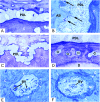
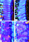
Mechanical testing of the periodontal ligament requires a practical experimental model. Bovine teeth are advantageous in terms of size and availability, but information is lacking as to the anatomy and histology of their periodontium. The aim of this study, therefore, was to characterize the anatomy and histology of the attachment apparatus in fully erupted bovine mandibular first molars. A total of 13 teeth were processed for the production of undecalcified ground sections and decalcified semi-thin sections, for NaOH maceration, and for polarized light microscopy. Histomorphometric measurements relevant to the mechanical behavior of the periodontal ligament included width, number, size and area fraction of blood vessels and fractal analysis of the two hard-soft tissue interfaces. The histological and histomorphometric analyses were performed at four different root depths and at six circumferential locations around the distal and mesial roots. The variety of techniques applied provided a comprehensive view of the tissue architecture of the bovine periodontal ligament. Marked regional variations were observed in width, surface geometry of the two bordering hard tissues (cementum and alveolar bone), structural organization of the principal periodontal ligament connective tissue fibers, size, number and numerical density of blood vessels in the periodontal ligament. No predictable pattern was observed, except for a statistically significant increase in the area fraction of blood vessels from apical to coronal. The periodontal ligament width was up to three times wider in bovine teeth than in human teeth. The fractal analyses were in agreement with the histological observations showing frequent signs of remodeling activity in the alveolar bone - a finding which may be related to the magnitude and direction of occlusal forces in ruminants. Although samples from the apical root portion are not suitable for biomechanical testing, all other levels in the buccal and lingual aspects of the mesial and distal roots may be considered. The bucco-mesial aspect of the distal root appears to be the most suitable location.
Figures


Similar articles
-
Periodontal ligament tissue reactions to trauma and gingival inflammation. An experimental study in the beagle dog.J Clin Periodontol. 1995 Oct;22(10):772-9. doi: 10.1111/j.1600-051x.1995.tb00260.x. J Clin Periodontol. 1995. PMID: 8682924
-
New attachment formation on teeth with a reduced but healthy periodontal ligament.J Clin Periodontol. 1985 Jan;12(1):51-60. doi: 10.1111/j.1600-051x.1985.tb01353.x. J Clin Periodontol. 1985. PMID: 3855871
-
Comparison of biomechanical properties of the incisor periodontal ligament among different species.Anat Rec. 1998 Apr;250(4):408-17. doi: 10.1002/(SICI)1097-0185(199804)250:4<408::AID-AR3>3.0.CO;2-T. Anat Rec. 1998. PMID: 9566530
-
Biomechanical pathways of dentoalveolar fibrous joints in health and disease.Periodontol 2000. 2020 Feb;82(1):238-256. doi: 10.1111/prd.12306. Periodontol 2000. 2020. PMID: 31850635 Review.
-
Developmental pathways of periodontal tissue regeneration: Developmental diversities of tooth morphogenesis do also map capacity of periodontal tissue regeneration?J Periodontal Res. 2019 Feb;54(1):10-26. doi: 10.1111/jre.12596. Epub 2018 Sep 12. J Periodontal Res. 2019. PMID: 30207395 Review.
Cited by
-
Challenges of Periodontal Tissue Engineering: Increasing Biomimicry through 3D Printing and Controlled Dynamic Environment.Nanomaterials (Basel). 2022 Nov 2;12(21):3878. doi: 10.3390/nano12213878. Nanomaterials (Basel). 2022. PMID: 36364654 Free PMC article. Review.
-
Bite force data suggests relationship between acrodont tooth implantation and strong bite force.PeerJ. 2020 Jun 30;8:e9468. doi: 10.7717/peerj.9468. eCollection 2020. PeerJ. 2020. PMID: 32656000 Free PMC article.
-
Influence of Micropatterning on Human Periodontal Ligament Cells' Behavior.Biophys J. 2018 Apr 24;114(8):1988-2000. doi: 10.1016/j.bpj.2018.02.041. Biophys J. 2018. PMID: 29694875 Free PMC article.
-
Three-dimensional morphometry of strained bovine periodontal ligament using synchrotron radiation-based tomography.J Anat. 2010 Aug;217(2):126-34. doi: 10.1111/j.1469-7580.2010.01250.x. Epub 2010 Jun 14. J Anat. 2010. PMID: 20557399 Free PMC article.
-
Dental ontogeny in extinct synapsids reveals a complex evolutionary history of the mammalian tooth attachment system.Proc Biol Sci. 2018 Nov 7;285(1890):20181792. doi: 10.1098/rspb.2018.1792. Proc Biol Sci. 2018. PMID: 30404877 Free PMC article.
References
-
- Ainamo J. Morphogenetic and functional characteristics of coronal cementum in bovine molars. Scand J Dent Res. 1970;78:378–386. - PubMed
-
- Andersson L, Malmgren B. The problem of dentoalveolar ankylosis and subsequent replacement resorption in the growing patient. Aust Endod J. 1999;25:57–61. - PubMed
-
- Beertsen W, McCulloch CA, Sodek J. The periodontal ligament: a unique, multifunctional connective tissue. Periodontol 2000. 1997;13:20–40. - PubMed
-
- Benatti BB, Silverio KG, Casati MZ, Sallum EA, Nociti FH., Jr Physiological features of periodontal regeneration and approaches for periodontal tissue engineering utilizing periodontal ligament cells. J Biosci Bioeng. 2007;103:1–6. - PubMed
-
- Berkovitz BK. Periodontal ligament: structural and clinical correlates. Dent Update. 2004;31:46–50. 52, 54. - PubMed
Publication types
MeSH terms
LinkOut - more resources
Full Text Sources
Other Literature Sources

