. 2008 Feb 27:8:22.
doi: 10.1186/1471-213X-8-22.

Expression of the Ladybird-like homeobox 2 transcription factor in the developing mouse testis and epididymis

Affiliations

Expression of the Ladybird-like homeobox 2 transcription factor in the developing mouse testis and epididymis

Vanessa Moisan et al. BMC Dev Biol. .

Abstract

Background: Homeoproteins are a class of transcription factors that are well-known regulators of organogenesis and cell differentiation in numerous tissues, including the male reproductive system. Indeed, a handful of homeoproteins have so far been identified in the testis and epididymis where a few were shown to play important developmental roles. Through a degenerate PCR approach aimed at identifying novel homeoproteins expressed in the male reproductive system, we have detected several homeoproteins most of which had never been described before in this tissue. One of these homeoproteins is Ladybird-like homeobox 2 (Lbx2), a homeobox factor mostly known to be expressed in the nervous system.

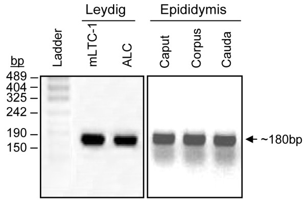
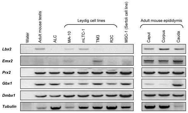
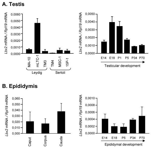
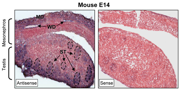
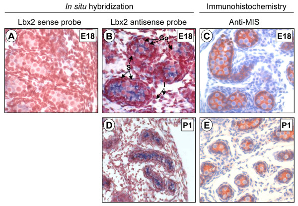
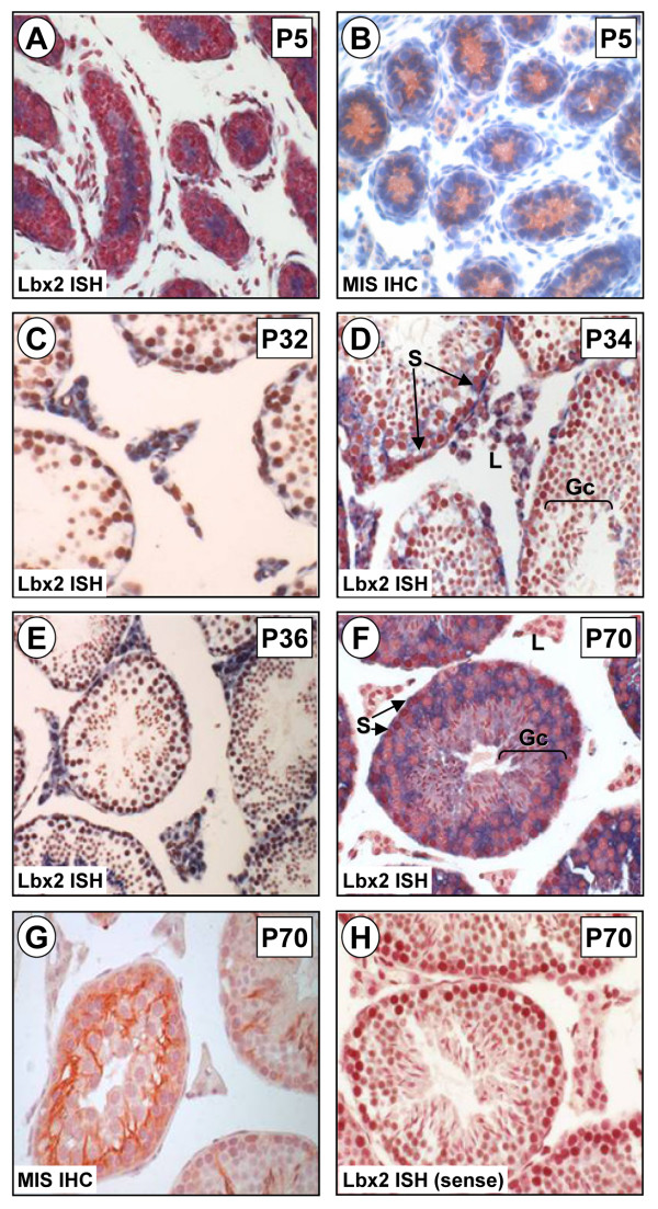
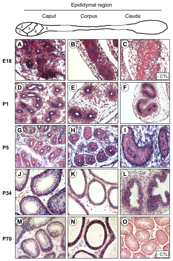
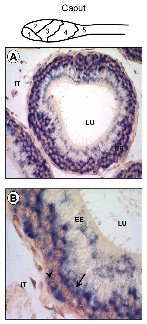
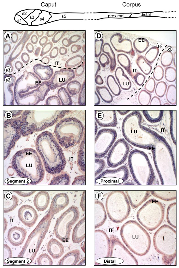
Results: To better define the expression profile of Lbx2 in the male reproductive system, we have performed in situ hybridization throughout testicular and epididymal development and into adulthood. Lbx2 expression was also confirmed by real time RT-PCR in those tissues and in several testicular and epididymal cell lines. In the epididymis, a highly segmented tissue, Lbx2 shows a regionalized expression profile, being more expressed in proximal segments of the caput epididymis than any other segment. In the testis, we found that Lbx2 is constitutively expressed at high levels in Sertoli cells. In interstitial cells, Lbx2 is weakly expressed during fetal and early postnatal life, highly expressed around P32-P36, and absent in adult animals. Finally, Lbx2 can also be detected in a population of germ cells in adults.

Conclusion: Altogether, our data suggest that the homeoprotein Lbx2 might be involved in the regulation of male reproductive system development and cell differentiation as well as in male epididymal segmentation.


Figures

Figure 1
Figure 1
Identification of homeobox factors in testicular Leydig cells and in mouse epididymis. A degenerate PCR was performed using cDNAs from mLTC-1 Leydig cell line, purified Leydig cells from adult rats (ALC) and from all three segments of the mouse epididymis and resulted in the amplification of a 180 bp fragment. The fragments were subcloned in pBluescript and sequenced to determine the nature of the homeoprotein (see Table 1).
Figure 2
Figure 2
Expression of five homeoproteins in the testis and epididymis. PCR reactions were performed using sequence-specific primers (see Table 2) for each of the indicated homeoproteins along with cDNAs from adult mouse testis, purified Leydig cells from adult rats (ALC), various Leydig cell lines (MA-10, mLTC-1, TM3, R2C), a Sertoli cell line (MSC-1), and the three regions of adult mouse epididymis (caput, corpus, cauda). The integrity and amount of cDNA used in the PCR assays was assessed by amplifying tubulin mRNA. No template: negative control.
Figure 3
Figure 3
Lbx2 is expressed in the testis (A) and epididymis (B). Quantitative real time PCR were performed with primers specific for Lbx2 cDNA as described in Methods using first strand cDNAs from Leydig cell lines (MA-10, mLTC-1, TM3), Sertoli cell lines (TM4, MSC-1, 15P-1), mouse testis at various developmental ages (E14, E18, P1, P5, P34 and P70), epididymal regions (caput, corpus, cauda) from adult mice, and mouse epididymis at different developmental stages as indicated. Results were corrected with the Rpl19 cDNA. Results are the mean of three individual experiments each performed in duplicate (± SEM).
Figure 4
Figure 4
Lbx2 is strongly expressed in mesonephric cells and in seminiferous tubules of the developing testis. Six μm paraffin sections of paraformaldehyde-fixed mouse developing urogenital ridge (E14) were probed with DIG-labeled antisense (left panel) and sense (right panel) Lbx2 cRNA probes in in situ hybridization experiments. Lbx2 mRNA was detected by immunostaining using an alkaline phosphatase-coupled anti-DIG antibody (appears as a blue-purplish staining). Tissues were counterstained with Neutral Red to visualize nuclei. Lbx2 mRNA was detected in cells of the Wolffian duct (WD), of the degenerating Müllerian duct (MD), and in the developing seminiferous tubules (ST, outlined by dotted lines). Magnification: 200×.
Figure 5
Figure 5
Lbx2 expression in embryonic an neonatal testis. Six μm paraffin sections of paraformaldehyde-fixed mouse testis were probed with DIG-labeled antisense (B and D) and sense (A) Lbx2 cRNA probes in in situ hybridization experiments. Lbx2 mRNA was detected by immunostaining using an alkaline phosphatase-coupled anti-DIG antibody (appears as a blue-purplish staining). Tissues were counterstained with Neutral Red to visualize nuclei. Lbx2 expression in the testis was assessed at E18 (B) and P1 (D). Immunohistochemistry for the Sertoli cell marker MIS was performed on testis sections at E18 (C) and P1 (E) as described in Methods and revealed using AEC (shows as a red-brownish staining). Expression of Lbx2 is evident within the cytoplasm of Sertoli cells by comparison with MIS staining. A weak but consistent Lbx2 staining is also observed in interstitial cells. Gonocytes are not labeled. No significant signal was detected with the sense probe (A). Go: gonocytes, S: Sertoli cells, I: interstitial cells. Magnifications: 200× (D, E), 400× (A-C).
Figure 6
Figure 6
Lbx2 expression in postnatal testis. In situ hybridization (ISH) experiments using DIG-labeled antisense (A, C, D, E, F) and sense (H) Lbx2 cRNA probes were performed on 6 μm paraffin sections of paraformaldehyde-fixed mouse testis. Lbx2 mRNA was detected by immunostaining using an alkaline phosphatase-coupled anti-DIG antibody (appears as a blue-purplish staining). Tissues were counterstained with Neutral Red to visualize nuclei. Lbx2 expression in the post-natal testis was assessed at P5 (A), P32 (C), P34 (D), P36 (E), and P70 (F). By immunohistochemistry (IHC), the Sertoli cell cytoplasm in P5 (B) and P70 (G) testis was labeled using an anti-MIS antiserum and revealed using AEC (shows as a red-brownish staining). Expression of Lbx2 is evident within the cytoplasm of Sertoli cells at all ages. In interstitial cells, a weak but consistent staining is observed at P5 (A) while a strong signal is detected at P32, P34, and P36 (C-E). At P70, some germ cells are also labeled for Lbx2 (F). No significant signal was detected with a sense probe as shown in the adult (H) section. Gc: germ cells, L: Leydig cells, S: Sertoli cells. Magnifications: 200× (A, B), 400× (C-H).
Figure 7
Figure 7
Lbx2 is strongly expressed throughout epididymal development. Top panel: schematic representation of the three regions of the epididymis and of the 5 segments of the caput. In situ hybridization experiments were performed on six μm paraffin sections of paraformaldehyde-fixed mouse epididymis using DIG-labeled antisense (A, B, D-N) and sense (C, O) Lbx2 cRNA probes. Lbx2 mRNA was detected by immunostaining using an alkaline phosphatase-coupled anti-DIG antibody (appears as a blue-purplish staining). Tissues were counterstained with Neutral Red to visualize nuclei. Lbx2 expression was assessed at different developmental stages, E18 (A, B), P1 (D-F), P5 (G-I), P34 (J-L), P70 (M, N), and in the three regions, caput (A, D, G, J, M), corpus (B, E, H, K, N), cauda (F, I, L) of the epididymis. No significant signal was detected with a sense probe as shown in the E18 (C) and adult (O) sections (- CTL). Magnifications: 100× (C, F, O); 200× (A, B, D, E, G, H, K, M); 400× (I, J, L, N).
Figure 8
Figure 8
Lbx2 is expressed in the principal and basal cells of the epididymal epithelium. Top panel: schematic representation of the caput epididymis and its 5 segments. A DIG-labeled antisense Lbx2 cRNA probe was used in in situ hybridization experiments on six μm paraffin sections of paraformaldehyde-fixed mouse adult epididymis. Lbx2 mRNA was detected by immunostaining using an alkaline phosphatase-coupled anti-DIG antibody (appears as a blue-purplish staining). Tissues were counterstained with Neutral Red to visualize nuclei. (A) 400× magnification of the segment 2 of the caput epididymis that reveals strong signal in the epididymal epithelium (EE). (B) The presence of Lbx2 can be observed in principal (arrow) and basal (arrowhead) cells using a 1000× magnification of (A). EE: epididymal epithelium; IT: interstitial compartment; LU; epididymal lumen.
Figure 9
Figure 9
Lbx2 is expressed in a segment-specific manner in the adult epididymis. Top panel: schematic representation of the five segments of the caput and the two regions of the corpus epididymis. Six μm paraffin sections of paraformaldehyde-fixed mouse epididymis were probed with a DIG-labeled antisense Lbx2 cRNA probe in in situ hybridization experiments. Lbx2 mRNA was detected by immunostaining using an alkaline phosphatase-coupled anti-DIG antibody (appears as a blue-purplish staining). Tissues were counterstained with Neutral Red to visualize nuclei. Caput (A, B, C) and corpus (D, E, F). (A) Localization of Lbx2 in segment 2 (s2) and 3 (s3) of the caput. The two segments are separated by a dotted line. (B) Magnification of segment 2 seen in A. (C) Magnification of segment 3 seen in A. (D) Lbx2 expression in the proximal (p) and distal (d) corpus. A dotted line separates the proximal and distal corpus. (E) Magnification of the proximal corpus seen in D. (F) Magnification of the distal corpus seen in (D). EE: epididymal epithelium; IT: interstitial compartment; LU: epididymal lumen. Magnifications: 100× (A, D); 200× (B, C, E, F).

Similar articles

Cited by

References

    1. Desplan C, Theis J, O'Farrell PH. The Drosophila developmental gene, engrailed, encodes a sequence-specific DNA binding activity. Nature. 1985;318:630–635. doi: 10.1038/318630a0. - DOI - PMC - PubMed
    1. Desplan C, Theis J, O'Farrell PH. The sequence specificity of homeodomain-DNA interaction. Cell. 1988;54:1081–1090. doi: 10.1016/0092-8674(88)90123-7. - DOI - PMC - PubMed
    1. Hoey T, Levine M. Divergent homeo box proteins recognize similar DNA sequences in Drosophila. Nature. 1988;332:858–861. doi: 10.1038/332858a0. - DOI - PubMed
    1. Bomgardner D, Hinton BT, Turner TT. Hox transcription factors may play a role in regulating segmental function of the adult epididymis. J Androl. 2001;22:527–531. - PubMed
    1. Lindsey JS, Wilkinson MF. Pem: a testosterone- and LH-regulated homeobox gene expressed in mouse Sertoli cells and epididymis. Dev Biol. 1996;179:471–484. doi: 10.1006/dbio.1996.0276. - DOI - PubMed

Publication types