Comparative Study
. 2008 Feb 27:8:69.
doi: 10.1186/1471-2148-8-69.

Molecular evolution of rDNA in early diverging Metazoa: first comparative analysis and phylogenetic application of complete SSU rRNA secondary structures in Porifera

Affiliations
Comparative Study

Molecular evolution of rDNA in early diverging Metazoa: first comparative analysis and phylogenetic application of complete SSU rRNA secondary structures in Porifera

Oliver Voigt et al. BMC Evol Biol. .

Abstract

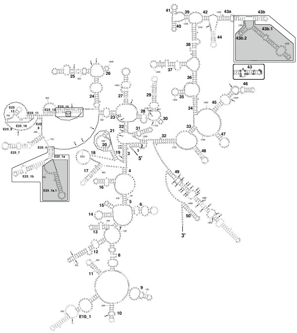
Background: The cytoplasmic ribosomal small subunit (SSU, 18S) ribosomal RNA (rRNA) is the most frequently-used gene for molecular phylogenetic studies. However, information regarding its secondary structure is neglected in most phylogenetic analyses. Incorporation of this information is essential in order to apply specific rRNA evolutionary models to overcome the problem of co-evolution of paired sites, which violates the basic assumption of the independent evolution of sites made by most phylogenetic methods. Information about secondary structure also supports the process of aligning rRNA sequences across taxa. Both aspects have been shown to increase the accuracy of phylogenetic reconstructions within various taxa.Here, we explore SSU rRNA secondary structures from the three extant classes of Phylum Porifera (Grant, 1836), a pivotal, but largely unresolved taxon of early branching Metazoa. This is the first phylogenetic study of poriferan SSU rRNA data to date that includes detailed comparative secondary structure information for all three sponge classes.

Results: We found base compositional and structural differences in SSU rRNA among Demospongiae, Hexactinellida (glass sponges) and Calcarea (calcareous sponges). We showed that analyses of primary rRNA sequences, including secondary structure-specific evolutionary models, in combination with reconstruction of the evolution of unusual structural features, reveal a substantial amount of additional information. Of special note was the finding that the gene tree topologies of marine haplosclerid demosponges, which are inconsistent with the current morphology-based classification, are supported by our reconstructed evolution of secondary structure features. Therefore, these features can provide alternative support for sequence-based topologies and give insights into the evolution of the molecule itself. To encourage and facilitate the application of rRNA models in phylogenetics of early metazoans, we present 52 SSU rRNA secondary structures over the taxonomic range of Porifera in a database, along with some basic tools for relevant format-conversion.

Conclusion: We demonstrated that sophisticated secondary structure analyses can increase the potential phylogenetic information of already available rDNA sequences currently accessible in databases and conclude that the importance of SSU rRNA secondary structure information for phylogenetic reconstruction is still generally underestimated, at least among certain early branching metazoans.


Figures

Figure 1
Figure 1
GC content against SSU rRNA fragment length. (Fragment corresponds to A. queenslandica positions 48–1896). A ca. 95% -fragment of SSU rRNA was used for analysis and only sequences with sequence information over the whole range of this fragment were considered (n = 123). Note that Farrea occa (Hexactinellida, [GenBank: AF159623]) is an incomplete potential pseudogene sequence.
Figure 2
Figure 2
SSU rRNA secondary structure for Calcarea. Sequence is given as 90% consensus with variable positions in black boxes. Lower case indicates deletions at the site for some sequences, according to the consensus level. Differences in helices between Calcaronea and Calcinea are in frames (Calcaronea = black, Calcinea = grey). Synapomorphies for each subclass are shown in boxes with the same color code. Primer positions are bold at the 5' and 3' end, respectively. Open circles instead of dots mark positions where mismatches occur in some sequences. Inset: Shortening and elongations in the boxed part of Helix E10_1 for two calcaronean sequences and one calcinean sequence.
Figure 3
Figure 3
SSU rRNA secondary structure of Acanthascus dawsoni [GenBank: AF100949] (Lyssacinosida, Rossellidae). Hexactinellid-specific helical insertions within E23_1 are shown in a box. Inset: Prediction of secondary structure insertions in E23_1 within other Hexactinellida. The insertions are predicted to form two helices in Hexasterophora (Lyssacinosida + Hexactinosida), and one helix in Amphidiscophora (Semperella schulzei). *Note that Farrea occa (AF159623) represents an (in other than the displayed part) incomplete, potential pseudogene molecule.
Figure 4
Figure 4
SSU rRNA secondary structure of the demosponge Amphimedon queenslandica (Haplosclerida). Nucleotides conserved in Demospongiae at the 90% level are shown in black, other nucleotides are in grey. Nucleotides at positions that are present in demosponges above the 90% consensus level but differ from A. queenslandica nucleotides are shown with an arrow pointing to their corresponding position. Specific insertions for A. queenslandica that are atypical for demosponges are displayed in shaded frames. Outlined frames highlight the regions of insertion within Haplosclerida that are displayed as sketches in Fig. 6. Inset: 90% consensus sequence and structure of partial helix 43 for 76 demosponges that do not belong to the marine haplosclerids.
Figure 5
Figure 5
Phylogeny inferred with PHASE. Nodes that differ from the topology published by Redmond et al. [41] are encircled. The boxed clades correspond to the excerpt displayed in Fig. 6. Support values are given at, or close to the corresponding node (values from analyses with PHASE/MrBayes; where the same support values were found in both analyses, only one number is shown; '<' = support values below 50; '-' = node not recovered in MrBayes analysis.). Monophyletic higher taxa are assigned.
Figure 6
Figure 6
Relationships of marine Haplosclerida (excerpt from larger phylogenetic analyses shown in Fig. 5) and evolution of extension regions. Sketches of predicted secondary structures for extensions and conserved flanking regions correspond to outlined boxes in Fig. 4. Asterisks mark nodes that were found in at least 96% of sampled trees after burn-in in both Bayesian analyses (PHASE and MrBayes, see Material and Methods for details); plus signs mark nodes that appeared in lower frequencies, but still above 84% in one, or both of the analyses. For each species, the family is shown below the sequence name.

References

    1. National Center for Biotechnology Information (GenBank) http://www.ncbi.nlm.nih.gov/
    1. Higgs PG. RNA secondary structure: physical and computational aspects. Q Rev Biophys. 2000;33:199–253. doi: 10.1017/S0033583500003620. - DOI - PubMed
    1. Green R, Noller HF. Ribosomes and translation. Annu Rev Biochem. 1997;66:679–716. doi: 10.1146/annurev.biochem.66.1.679. - DOI - PubMed
    1. Kjer KM. Use of rRNA secondary structure in phylogenetic studies to identify homologous positions: an example of alignment and data presentation from the frogs. Mol Phylogenet Evol. 1995;4:314–330. doi: 10.1006/mpev.1995.1028. - DOI - PubMed
    1. Hickson RE, Simon C, Perrey SW. The performance of several multiple-sequence alignment programs in relation to secondary-structure features for an rRNA sequence. Mol Biol Evol. 2000;17:530–539. - PubMed

Publication types

LinkOut - more resources