

Splice mutation in the iron-sulfur cluster scaffold protein ISCU causes myopathy with exercise intolerance
- PMID: 18304497
- PMCID: PMC2427212
- DOI: 10.1016/j.ajhg.2007.12.012
Splice mutation in the iron-sulfur cluster scaffold protein ISCU causes myopathy with exercise intolerance
Abstract
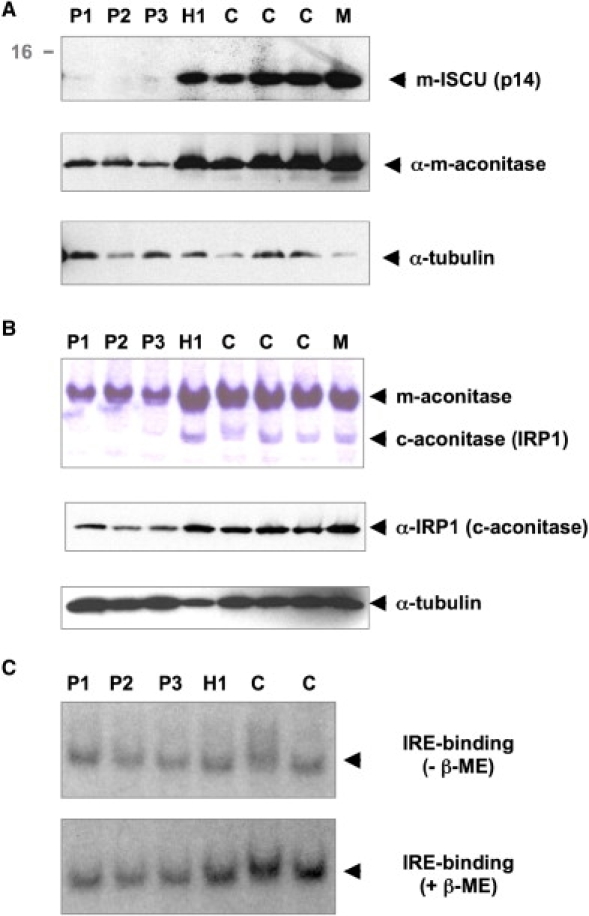
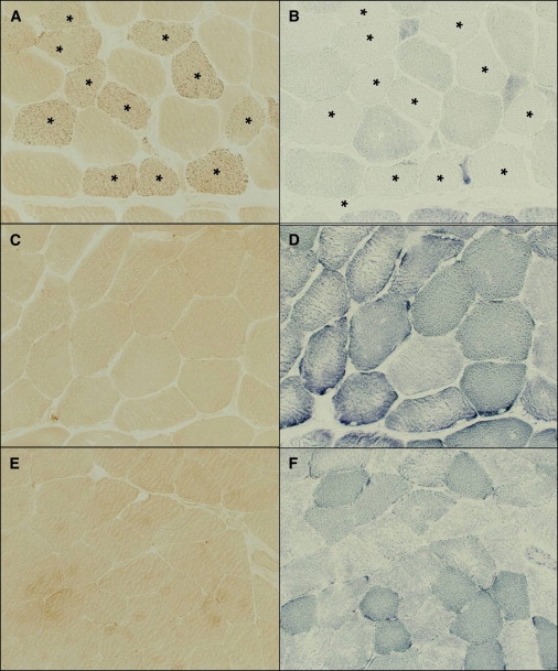
A myopathy with severe exercise intolerance and myoglobinuria has been described in patients from northern Sweden, with associated deficiencies of succinate dehydrogenase and aconitase in skeletal muscle. We identified the gene for the iron-sulfur cluster scaffold protein ISCU as a candidate within a region of shared homozygosity among patients with this disease. We found a single mutation in ISCU that likely strengthens a weak splice acceptor site, with consequent exon retention. A marked reduction of ISCU mRNA and mitochondrial ISCU protein in patient muscle was associated with a decrease in the iron regulatory protein IRP1 and intracellular iron overload in skeletal muscle, consistent with a muscle-specific alteration of iron homeostasis in this disease. ISCU interacts with the Friedreich ataxia gene product frataxin in iron-sulfur cluster biosynthesis. Our results therefore extend the range of known human diseases that are caused by defects in iron-sulfur cluster biogenesis.
Figures
Similar articles
-
Clinical manifestation and a new ISCU mutation in iron-sulphur cluster deficiency myopathy.Brain. 2009 Aug;132(Pt 8):2170-9. doi: 10.1093/brain/awp152. Epub 2009 Jun 30. Brain. 2009. PMID: 19567699
-
The presence of multiple cellular defects associated with a novel G50E iron-sulfur cluster scaffold protein (ISCU) mutation leads to development of mitochondrial myopathy.J Biol Chem. 2014 Apr 11;289(15):10359-10377. doi: 10.1074/jbc.M113.526665. Epub 2014 Feb 26. J Biol Chem. 2014. PMID: 24573684 Free PMC article.
-
A novel de novo dominant mutation in ISCU associated with mitochondrial myopathy.J Med Genet. 2017 Dec;54(12):815-824. doi: 10.1136/jmedgenet-2017-104822. Epub 2017 Oct 27. J Med Genet. 2017. PMID: 29079705 Free PMC article.
-
Human iron-sulfur cluster assembly, cellular iron homeostasis, and disease.Biochemistry. 2010 Jun 22;49(24):4945-56. doi: 10.1021/bi1004798. Biochemistry. 2010. PMID: 20481466 Free PMC article. Review.
-
[Mitochondrial aconitase].Nihon Rinsho. 2002 Apr;60 Suppl 4:133-4. Nihon Rinsho. 2002. PMID: 12013835 Review. Japanese. No abstract available.
Cited by
-
Downregulation of Iron-Sulfur Cluster Biogenesis May Contribute to Hyperglycemia-Mediated Diabetic Peripheral Neuropathy in Murine Models.Antioxidants (Basel). 2024 Aug 26;13(9):1036. doi: 10.3390/antiox13091036. Antioxidants (Basel). 2024. PMID: 39334695 Free PMC article.
-
Rats with a Human Mutation of NFU1 Develop Pulmonary Hypertension.Am J Respir Cell Mol Biol. 2020 Feb;62(2):231-242. doi: 10.1165/rcmb.2019-0065OC. Am J Respir Cell Mol Biol. 2020. PMID: 31461310 Free PMC article.
-
The Impact of Iron Overload and Ferroptosis on Reproductive Disorders in Humans: Implications for Preeclampsia.Int J Mol Sci. 2019 Jul 4;20(13):3283. doi: 10.3390/ijms20133283. Int J Mol Sci. 2019. PMID: 31277367 Free PMC article. Review.
-
FDX2 and ISCU Gene Variations Lead to Rhabdomyolysis With Distinct Severity and Iron Regulation.Neurol Genet. 2022 Jan 19;8(1):e648. doi: 10.1212/NXG.0000000000000648. eCollection 2022 Feb. Neurol Genet. 2022. PMID: 35079622 Free PMC article.
-
Iron-dependent functions of mitochondria--relation to neurodegeneration.J Neural Transm (Vienna). 2011 Mar;118(3):349-59. doi: 10.1007/s00702-010-0503-7. Epub 2010 Dec 15. J Neural Transm (Vienna). 2011. PMID: 21161302 Review.
References
-
- Linderholm H., Muller R., Ringqvist T., Sornas R. Hereditary abnormal muscle metabolism with hyperkinetic circulation during exercise. Acta Med. Scand. 1969;185:153–166. - PubMed
-
- Linderholm H., Essen-Gustavsson B., Thornell L.E. Low succinate dehydrogenase (SDH) activity in a patient with a hereditary myopathy with paroxysmal myoglobinuria. J. Intern. Med. 1990;228:43–52. - PubMed
-
- Haller R.G., Henriksson K.G., Jorfeldt L., Hultman E., Wibom R., Sahlin K., Areskog N.H., Gunder M., Ayyad K., Blomqvist C.G. Deficiency of skeletal muscle succinate dehydrogenase and aconitase. Pathophysiology of exercise in a novel human muscle oxidative defect. J. Clin. Invest. 1991;88:1197–1206. - PMC - PubMed
Publication types
MeSH terms
Substances
Associated data
- Actions
- Actions
Grants and funding
LinkOut - more resources
Full Text Sources
Medical
Molecular Biology Databases
