. 2008 Mar;109(1-2):150-7.
doi: 10.1016/j.jsbmb.2008.01.002. Epub 2008 Jan 19.

Differential recruitment of glucocorticoid receptor phospho-isoforms to glucocorticoid-induced genes

Affiliations

Differential recruitment of glucocorticoid receptor phospho-isoforms to glucocorticoid-induced genes

Raymond D Blind et al. J Steroid Biochem Mol Biol. 2008 Mar.

Abstract

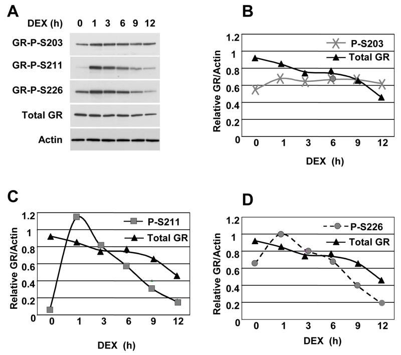
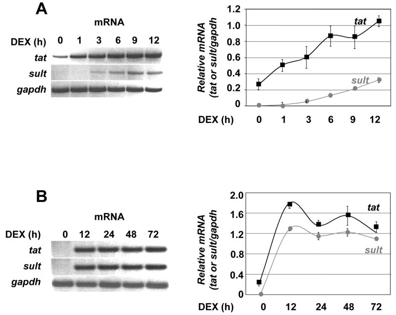
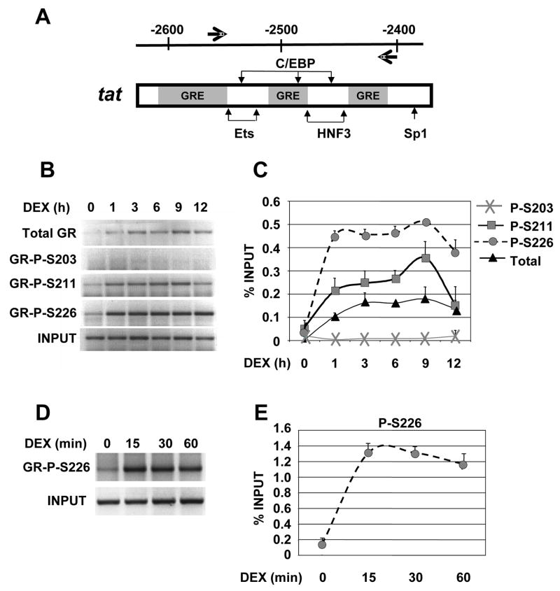
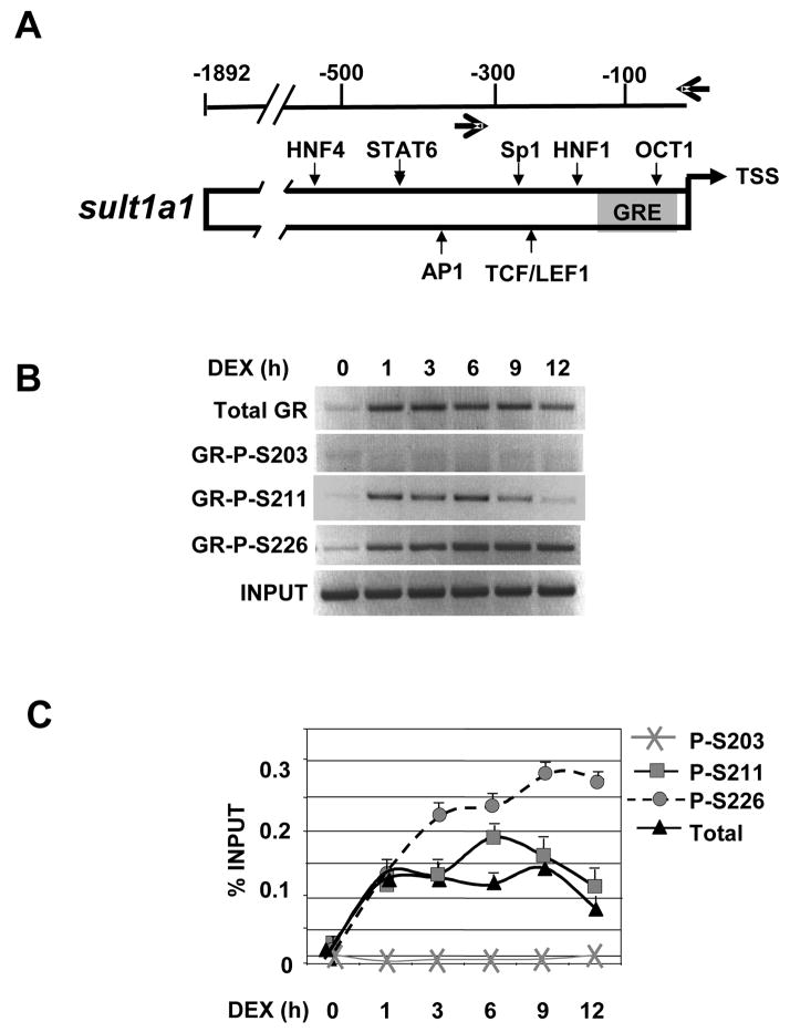
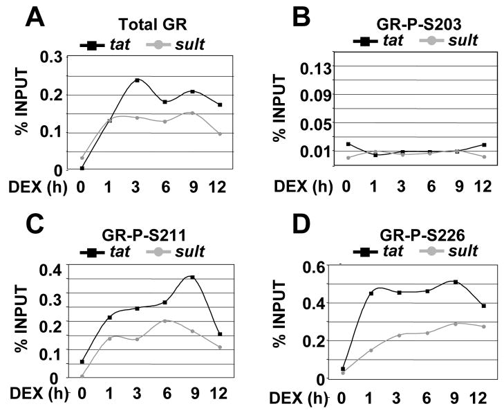
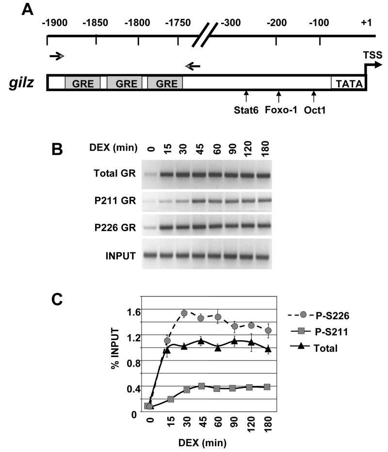
The human glucocorticoid receptor (GR) is phosphorylated on its N-terminus at three major sites (S203, S211 and S226) within activation function 1 (AF1). Although GR has been shown to assemble at glucocorticoid responsive elements (GREs) in the presence of hormone, the timing and specificity of GR phospho-isoform recruitment to receptor target genes has not been established. Using chromatin immunoprecipitation (ChIP) and GR phosphorylation site-specific antibodies, we examined GR phospho-isoform recruitment to several glucocorticoid-induced genes including tyrosine aminotransferase (tat) and sulfonyltransferase-1A1 (sult) in rat hepatoma cells, and the glucocorticoid-induced leucine zipper (gilz) gene in human U2OS cells. GR P-S211 and GR P-S226 isoforms were efficiently recruited to the tat, sult and gilz GREs in a hormone-dependent manner. In contrast, the GR P-S203 isoform displayed no significant recruitment to any GREs of the genes analyzed, consistent with its lack of nuclear accumulation. Interestingly, the kinetics of GR P-S211 and GR P-S226 recruitment differed among genes. Our findings indicate that GR phospho-isoforms selectively occupy GR target genes, and suggests gene specific requirements for GR phosphorylation in receptor-dependent transcriptional activation.


Figures

Figure 1
Figure 1. Kinetics of GR phosphorylation in rat hepatoma cells in response to Dex
A) Rat hepatoma cells were treated with ethanol (−) or Dex (100 nM) for the times indicated. Whole cell lysates were prepared, normalized and analyzed by immunoblotting with GR P-S203, GR P-S203, GR P-S226, total GR and actin antibodies. B–D) Quantitative analysis of total GR and phospho-GR immunoblots in panel A normalized to actin. The data shown are from a single experiment that is representative of at least three independent experiments.
Figure 2
Figure 2. Induction of tyrosine aminotransferase and sulfonyl transferase 1A1 by GR
A) Rat hepatoma cells were treated with 100 nM Dexamethasone (Dex) over a 12 hour period. Total RNA was harvested, reverse-transcribed, and subjected to PCR with primer pairs to indicated genes, with the gapdh mRNA used as an internal control. The PCR products were resolved on agarose gels, visualized by ethidium bromide staining and quantified relative to the gapdh control. B) RT-PCR done as in (A) over 72 hours. Results are from three independent experiments, each done in duplicate, with error bars representing standard deviation.
Figure 3
Figure 3. GR phospho-isoform recruitment to tyrosine aminotransferase (tat)
A) Schematic depiction of the tat regulatory regions. GR binding sites (GREs) are shown as gray boxes. The small vertical arrows show additional transcription factor binding sites. The large horizontal arrows represent the primers pairs used to amplify the region that contain the GREs. B) Phospho-GR recruitment to tat over 12 h. Rat hepatoma cells were treated with vehicle or Dex for the times indicated and ChIP were performed with antibodies against total GR, P-S203, P-S211, P-S226 and GR binding sites were amplified by PCR using tat-specific primer pairs and the PCR products resolved on agarose gels, visualized by ethidium bromide staining and quantified relative to input using NIH Image. C) The signal of the PCR product representing 0.5% input was arbitrarily set as 1. Data were averaged from three independent ChIP assays. The error bar represents S.D. D–E) GR P-S226 recruitment to tat over 60 min. Rat hepatoma cells were treated with vehicle or Dex for 0, 15, 30 and 60 min and ChIP were performed and quantitated as described above.
Figure 4
Figure 4. GR phospho-isoform recruitment to sulfonyl transferase 1A1 (sult)
A) Schematic depiction of the sult regulatory region. The single promoter proximal GR binding site is shown as a gray box. The small vertical arrows show other transcription factor binding sites. The large horizontal arrows depict the primers pairs used to amplify the region that flank the GRE. B–C) Phospho-GR recruitment to sult over 12 h. Rat hepatoma cells were treated with vehicle or Dex for the times indicated and ChIP were performed with antibodies against total GR, P-S203, P-S211, P-S226 and GR binding sites were amplified by PCR using sult -specific primer pairs and the PCR products resolved, stained and quantified as in Figure 3.
Figure 5
Figure 5. GR phospho-isoforms are differentially recruited to the tat and sult GREs
A) Total GR B) P-S203 C) P-S211 and D) P-S226 recruitment to tat and sult over 12 h plotted from Figures 3 and 4.
Figure 6
Figure 6. GR phospho-isoform recruitment to the gilz regulatory region
A) Schematic depiction of the gilz regulatory region. The GR binding sites (GREs) are shown as gray boxes. The small vertical arrows show other transcription factor binding sites. The horizontal arrows depict the primers pairs used to amplify the regions encompassing GREs. B–C) Phospho-GR recruitment to gilz over 180 min. U2OS-hGR cells were treated with vehicle or Dex for the times indicated and ChIP were performed with antibodies against total GR, P-S211, P-S226 and GR binding sites were amplified by PCR using gilz-specific primer pairs and the PCR products resolved, stained and quantified as in Figure 3.

References

    1. Buckingham JC. Glucocorticoids: exemplars of multi-tasking. Br J Pharmacol. 2006;147(Suppl 1):S258–68. - PMC - PubMed
    1. Yudt MR, Cidlowski JA. The glucocorticoid receptor: coding a diversity of proteins and responses through a single gene. Mol Endocrinol. 2002;16(8):1719–26. - PubMed
    1. Picard D. Chaperoning steroid hormone action. Trends Endocrinol Metab. 2006;17(6):229–35. - PubMed
    1. Yamamoto KR. Multilayered control of intracellular receptor function. Harvey Lect. 1995;91:1–19. - PubMed
    1. Kumar R, Thompson EB. Gene regulation by the glucocorticoid receptor: structure:function relationship. J Steroid Biochem Mol Biol. 2005;94(5):383–94. - PubMed

Publication types

MeSH terms