Nonhelical leash and alpha-helical structures determine the potency of a peptide antagonist of human T-cell leukemia virus entry
- PMID: 18305034
- PMCID: PMC2346759
- DOI: 10.1128/JVI.02458-07
Nonhelical leash and alpha-helical structures determine the potency of a peptide antagonist of human T-cell leukemia virus entry
Abstract
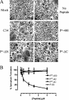
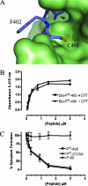
Viral fusion proteins mediate the entry of enveloped viral particles into cells by inducing fusion of the viral and target cell membranes. Activated fusion proteins undergo a cascade of conformational transitions and ultimately resolve into a compact trimer of hairpins or six-helix bundle structure, which pulls the interacting membranes together to promote lipid mixing. Significantly, synthetic peptides based on a C-terminal region of the trimer of hairpins are potent inhibitors of membrane fusion and viral entry, and such peptides are typically extensively alpha-helical. In contrast, an atypical peptide inhibitor of human T-cell leukemia virus (HTLV) includes alpha-helical and nonhelical leash segments. We demonstrate that both the C helix and C-terminal leash are critical to the inhibitory activities of these peptides. Amino acid side chains in the leash and C helix extend into deep hydrophobic pockets at the membrane-proximal end of the HTLV type 1 (HTLV-1) coiled coil, and these contacts are necessary for potent antagonism of membrane fusion. In addition, a single amino acid substitution within the inhibitory peptide improves peptide interaction with the core coiled coil and yields a peptide with enhanced potency. We suggest that the deep pockets on the coiled coil are ideal targets for small-molecule inhibitors of HTLV-1 entry into cells. Moreover, the extended nature of the HTLV-1-inhibitory peptide suggests that such peptides may be intrinsically amenable to modifications designed to improve inhibitory activity. Finally, we propose that leash-like mimetic peptides may be of value as entry inhibitors for other clinically important viral infections.
Figures


Similar articles
-
Highly specific inhibition of leukaemia virus membrane fusion by interaction of peptide antagonists with a conserved region of the coiled coil of envelope.Retrovirology. 2008 Aug 4;5:70. doi: 10.1186/1742-4690-5-70. Retrovirology. 2008. PMID: 18680566 Free PMC article.
-
An antiviral peptide targets a coiled-coil domain of the human T-cell leukemia virus envelope glycoprotein.J Virol. 2003 Mar;77(5):3281-90. doi: 10.1128/jvi.77.5.3281-3290.2003. J Virol. 2003. PMID: 12584351 Free PMC article.
-
Basic residues are critical to the activity of peptide inhibitors of human T cell leukemia virus type 1 entry.J Biol Chem. 2009 Mar 6;284(10):6575-84. doi: 10.1074/jbc.M806725200. Epub 2008 Dec 29. J Biol Chem. 2009. PMID: 19114713
-
Peptide-Based Dual HIV and Coronavirus Entry Inhibitors.Adv Exp Med Biol. 2022;1366:87-100. doi: 10.1007/978-981-16-8702-0_6. Adv Exp Med Biol. 2022. PMID: 35412136 Review.
-
Peptide entry inhibitors of enveloped viruses: the importance of interfacial hydrophobicity.Biochim Biophys Acta. 2014 Sep;1838(9):2180-97. doi: 10.1016/j.bbamem.2014.04.015. Epub 2014 Apr 26. Biochim Biophys Acta. 2014. PMID: 24780375 Free PMC article. Review.
Cited by
-
Charge-surrounded pockets and electrostatic interactions with small ions modulate the activity of retroviral fusion proteins.PLoS Pathog. 2011 Feb 3;7(2):e1001268. doi: 10.1371/journal.ppat.1001268. PLoS Pathog. 2011. PMID: 21304939 Free PMC article.
-
Cellular Factors Involved in HTLV-1 Entry and Pathogenicit.Front Microbiol. 2012 Jun 21;3:222. doi: 10.3389/fmicb.2012.00222. eCollection 2012. Front Microbiol. 2012. PMID: 22737146 Free PMC article.
-
Highly specific inhibition of leukaemia virus membrane fusion by interaction of peptide antagonists with a conserved region of the coiled coil of envelope.Retrovirology. 2008 Aug 4;5:70. doi: 10.1186/1742-4690-5-70. Retrovirology. 2008. PMID: 18680566 Free PMC article.
-
Current State of Therapeutics for HTLV-1.Viruses. 2024 Oct 15;16(10):1616. doi: 10.3390/v16101616. Viruses. 2024. PMID: 39459949 Free PMC article. Review.
-
Capturing a fusion intermediate of influenza hemagglutinin with a cholesterol-conjugated peptide, a new antiviral strategy for influenza virus.J Biol Chem. 2011 Dec 9;286(49):42141-42149. doi: 10.1074/jbc.M111.254243. Epub 2011 Oct 12. J Biol Chem. 2011. PMID: 21994935 Free PMC article.
References
-
- Chan, D. C., D. Fass, J. M. Berger, and P. S. Kim. 1997. Core structure of gp41 from the HIV envelope glycoprotein. Cell 89263-273. - PubMed
-
- Connor, R. I., B. K. Chen, S. Choe, and N. R. Landau. 1995. Vpr is required for efficient replication of human immunodeficiency virus type-1 in mononuclear phagocytes. Virology 206935-944. - PubMed
Publication types
MeSH terms
Substances
LinkOut - more resources
Full Text Sources

