

Individual crypt genetic heterogeneity and the origin of metaplastic glandular epithelium in human Barrett's oesophagus
- PMID: 18305067
- PMCID: PMC2564832
- DOI: 10.1136/gut.2007.143339
Individual crypt genetic heterogeneity and the origin of metaplastic glandular epithelium in human Barrett's oesophagus
Abstract
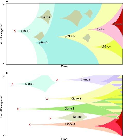
Objectives: Current models of clonal expansion in human Barrett's oesophagus are based upon heterogenous, flow-purified biopsy analysis taken at multiple segment levels. Detection of identical mutation fingerprints from these biopsy samples led to the proposal that a mutated clone with a selective advantage can clonally expand to fill an entire Barrett's segment at the expense of competing clones (selective sweep to fixation model). We aimed to assess clonality at a much higher resolution by microdissecting and genetically analysing individual crypts. The histogenesis of Barrett's metaplasia and neo-squamous islands has never been demonstrated. We investigated the oesophageal gland squamous ducts as the source of both epithelial sub-types.
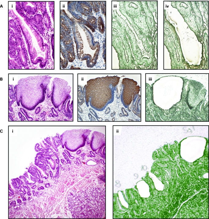
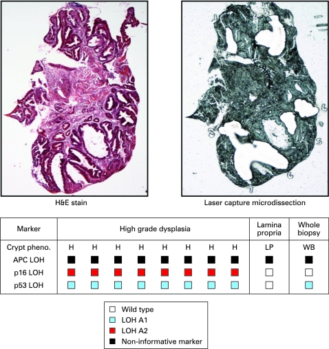
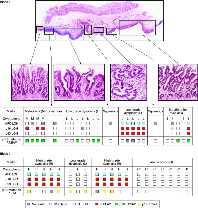
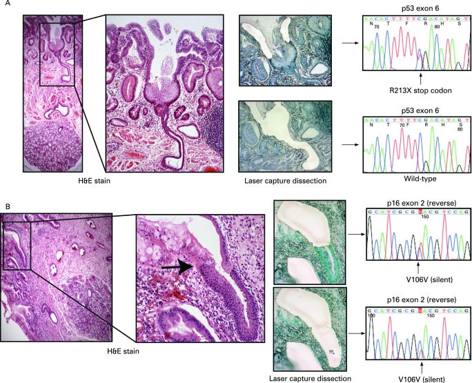
Methods: Individual crypts across Barrett's biopsy and oesophagectomy blocks were dissected. Determination of tumour suppressor gene loss of heterozygosity patterns, p16 and p53 point mutations were carried out on a crypt-by-crypt basis. Cases of contiguous neo-squamous islands and columnar metaplasia with oesophageal squamous ducts were identified. Tissues were isolated by laser capture microdissection and genetically analysed.
Results: Individual crypt dissection revealed mutation patterns that were masked in whole biopsy analysis. Dissection across oesophagectomy specimens demonstrated marked clonal heterogeneity, with multiple independent clones present. We identified a p16 point mutation arising in the squamous epithelium of the oesophageal gland duct, which was also present in a contiguous metaplastic crypt, whereas neo-squamous islands arising from squamous ducts were wild-type with respect to surrounding Barrett's dysplasia.
Conclusions: By studying clonality at the crypt level we demonstrate that Barrett's heterogeneity arises from multiple independent clones, in contrast to the selective sweep to fixation model of clonal expansion previously described. We suggest that the squamous gland ducts situated throughout the oesophagus are the source of a progenitor cell that may be susceptible to gene mutation resulting in conversion to Barrett's metaplastic epithelium. Additionally, these data suggest that wild-type ducts may be the source of neo-squamous islands.
Conflict of interest statement
Figures
Comment in
-
Dissecting out the genetic origins of Barrett's oesophagus.Gut. 2008 Aug;57(8):1033-4. doi: 10.1136/gut.2008.151530. Gut. 2008. PMID: 18628369 No abstract available.
Similar articles
-
Barrett's metaplasia glands are clonal, contain multiple stem cells and share a common squamous progenitor.Gut. 2012 Oct;61(10):1380-9. doi: 10.1136/gutjnl-2011-301174. Epub 2011 Dec 26. Gut. 2012. PMID: 22200839
-
On the histogenesis of Barrett's oesophagus and its associated squamous islands: a three-dimensional study of their morphological relationship with native oesophageal gland ducts.J Pathol. 2005 Aug;206(4):388-94. doi: 10.1002/path.1804. J Pathol. 2005. PMID: 15926200
-
K-ras point mutations are rare events in premalignant forms of Barrett's oesophagus.Eur J Gastroenterol Hepatol. 1996 Aug;8(8):799-804. Eur J Gastroenterol Hepatol. 1996. PMID: 8864678
-
In brief: the (molecular) pathogenesis of Barrett's oesophagus.J Pathol. 2014 Mar;232(4):383-5. doi: 10.1002/path.4300. J Pathol. 2014. PMID: 24254998 Review.
-
Cellular origin of Barrett's metaplasia and oesophageal stem cells.Biochem Soc Trans. 2010 Apr;38(2):370-3. doi: 10.1042/BST0380370. Biochem Soc Trans. 2010. PMID: 20298185 Review.
Cited by
-
Genetic progression of Barrett's oesophagus to oesophageal adenocarcinoma.Br J Cancer. 2016 Aug 9;115(4):403-10. doi: 10.1038/bjc.2016.219. Epub 2016 Jul 21. Br J Cancer. 2016. PMID: 27441494 Free PMC article. Review.
-
Epithelial cell plasticity: breaking boundaries and changing landscapes.EMBO Rep. 2021 Jul 5;22(7):e51921. doi: 10.15252/embr.202051921. Epub 2021 Jun 6. EMBO Rep. 2021. PMID: 34096150 Free PMC article. Review.
-
Ductal metaplasia in oesophageal submucosal glands is associated with inflammation and oesophageal adenocarcinoma.Histopathology. 2015 Dec;67(6):771-82. doi: 10.1111/his.12707. Epub 2015 Jun 4. Histopathology. 2015. PMID: 25847432 Free PMC article.
-
Clonal evolution in cancer.Nature. 2012 Jan 18;481(7381):306-13. doi: 10.1038/nature10762. Nature. 2012. PMID: 22258609 Free PMC article. Review.
-
Mystery Behind Barrett's Esophagus: The Origin and Malignant Transformation of Esophageal Adenocarcinoma.Glob Med Genet. 2022 Dec 21;9(4):287-289. doi: 10.1055/s-0042-1758764. eCollection 2022 Dec. Glob Med Genet. 2022. PMID: 36567952 Free PMC article. No abstract available.
References
-
- Lagergren J, Bergstrom R, Lindgren A, et al. Symptomatic gastroesophageal reflux as a risk factor for esophageal adenocarcinoma. N Engl J Med 1999;340:825–31 - PubMed
-
- Wong DJ, Paulson TG, Prevo LJ, et al. p16(INK4a) lesions are common, early abnormalities that undergo clonal expansion in Barrett’s metaplastic epithelium. Cancer Res 2001;61:8284–9 - PubMed
Publication types
MeSH terms
Grants and funding
LinkOut - more resources
Full Text Sources
Other Literature Sources
Research Materials
Miscellaneous