. 2008 May;19(5):891-903.
doi: 10.1681/ASN.2006111282. Epub 2008 Feb 27.

SIX2 and BMP4 mutations associate with anomalous kidney development

Affiliations

SIX2 and BMP4 mutations associate with anomalous kidney development

Stefanie Weber et al. J Am Soc Nephrol. 2008 May.

Abstract

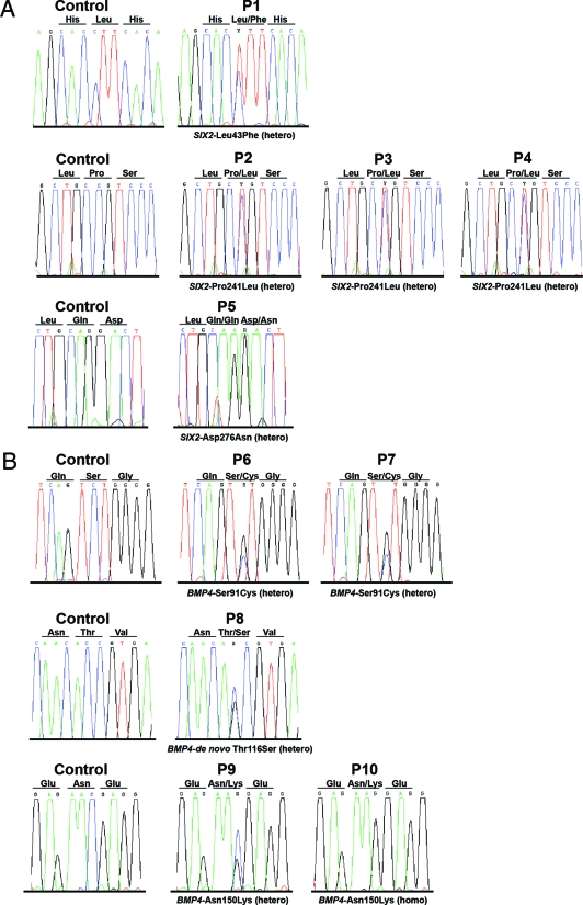
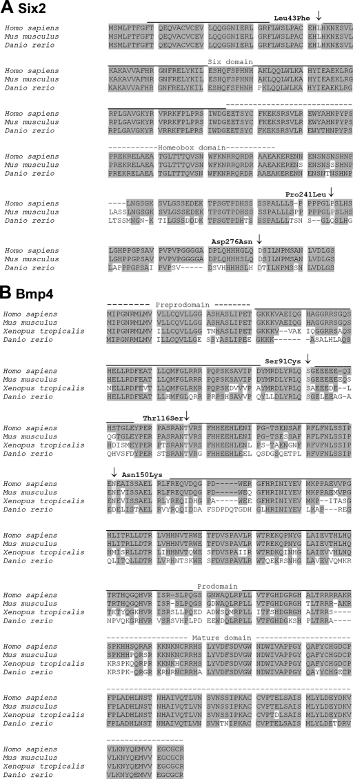
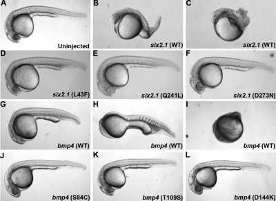
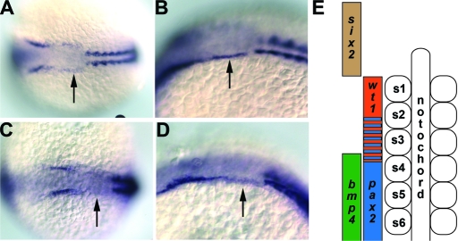
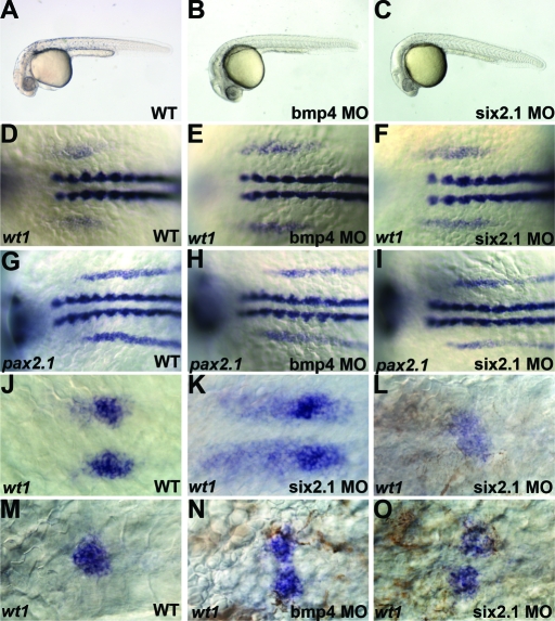
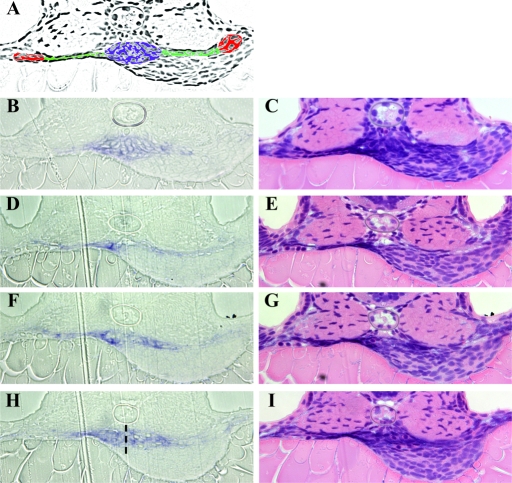
Renal hypodysplasia (RHD) is characterized by reduced kidney size and/or maldevelopment of the renal tissue following abnormal organogenesis. Mutations in renal developmental genes have been identified in a subset of affected individuals. Here, we report the first mutations in BMP4 and SIX2 identified in patients with RHD. We detected 3 BMP4 mutations in 5 RHD patients, and 3 SIX2 mutations in 5 different RHD patients. Overexpression assays in zebrafish demonstrated that these mutations affect the function of Bmp4 and Six2 in vivo. Overexpression of zebrafish six2.1 and bmp4 resulted in dorsalization and ventralization, respectively, suggesting opposing roles in mesendoderm formation. When mutant constructs containing the identified human mutations were overexpressed instead, these effects were attenuated. Morpholino knockdown of bmp4 and six2.1 affected glomerulogenesis, suggesting specific roles for these genes in the formation of the pronephros. In summary, these studies implicate conserved roles for Six2 and Bmp4 in the development of the renal system. Defects in these proteins could affect kidney development at multiple stages, leading to the congenital anomalies observed in patients with RHD.


Figures

Figure 1.
Figure 1.
Human mutations identified in SIX2 and BMP4. SIX2 (A) and BMP4 (B) DNA sequencing results are shown for all index patients and healthy controls.
Figure 2.
Figure 2.
Six2 and Bmp4 sequence alignments. (A) Alignment of Six2 amino acid (AA) sequences depicting the AA sites affected by the missense mutations identified in this study (Leu43Phe, Pro241Leu, Asp276Asn). Zebrafish Six2.1 shares 79% and 78% identity and 82% and 83% similarity to human and mouse Six2, respectively. GenBank accession numbers are as follows: Homo sapiens AAF69031, Mus musculus AAH68021, Danio rerio BAB40699. (B) Alignment of Bmp4 AA sequences demonstrating the identified mutations (Ser91Cys, Thr116Ser, Asn150Lys). Zebrafish Bmp4 shares 69%, 69%, and 68% identity and 80%, 80%, and 81% similarity to human, mouse, and frog Bmp4, respectively. GenBank accession numbers are as follows: Homo sapiens AAH20546, Mus musculus AAH34053, Xenopus tropicans AAY90071, Danio rerio AAH78423.
Figure 3.
Figure 3.
Six2 and Bmp4 expression in human fetal kidney. Immunohistochemistry for Bmp4 and Six2 in first-trimester human kidneys. All sections counterstained with hematoxylin, positive immunohistochemical signal is brown. (A-C) High power views of nephrogenic zone from 9 wk gestation kidney. (A) Branched ureteric bud tips (u), adjacent to condensing mesenchyme (cm), with deeper S-shaped body (s) and glomerular precursor (gp). (B) Only occasional cells positive for Bmp4 in mesenchyme (note light brown color within tubules in bottom right is a false positive caused by incomplete quenching of endogenous peroxidase). (C) Strongly positive signal for Six2 in uninduced mesenchyme and parts of condensed mesenchyme, ureteric buds are negative. (D) Sections through cortex of 12-wk gestation kidney (again note endogenous peroxidase). (E) Bmp4 expression is not consistently detected in the cortex at this stage. (F) Six2 is expressed in a rim of outer mesenchyme in the nephrogenic cortex. (G-J) Sections from a 12-wk gestation kidney including the medulla. (I) Higher power view of the deep medulla from panel G. (J) Higher power view of the deep medulla from panel H. Strongly positive expression of both Bmp4 and Six2 in large proximal ureteric branches in the medulla, with negative surrounding loose connective tissue. Bar = 20 μm in panels A through C, I, and J; 100 μm in D through H.
Figure 4.
Figure 4.
bmp4 and six2 overexpression phenotypes at 24 h. Lateral views of wild-type and RNA-injected zebrafish embryos at 24 h postfertilization. (A) Wild-type embryo (WT). (B-C) Embryos injected with 100 pg six2.1 RNA demonstrating class I to III (B) and class IV (C) dorsalized phenotypes. (D-F) Embryos injected with 100 pg of mutated six2.1 RNA: (D) six2 L43F, (E) six2 Q241L, (F) six2 D273N. (G-I) Embryos injected with 25 pg bmp4 RNA demonstrating class II (G), class III (H), and class IV (I) ventralized phenotypes. (J-L) Embryos injected with 25 pg of mutated bmp4 RNA (J), bmp4 S84C (K), bmp4 T109S (L), and bmp4 (D144K). See Table 2 for further description of ventralized and dorsalized classes.
Figure 5.
Figure 5.
Expression of bmp4 and six2.1. (A) Dorsal view of six2.1 RNA expression in a 10-somite stage embryo. Note the expression that extends posterior from the darker stained otic placode to the first somite (arrow). (B) Lateral view of the same embryo in (A). (C) Dorsal view of six2.1 RNA expression in a 14-somite stage embryo. Expression between the darker stained otic placode and the first somite is weaker than at 10 somites (arrow). (D) Lateral view of the same embryo in panel C. Anterior is to the left in panels A through D. Arrows denote the expression of six2.1 that extends to the first somite. Costaining for α-tropomyosin is used to indicate somites. (E) Diagram adapted from Serluca et al. depicting expression domains of genes used in this study. Anterior somites are numbered from anterior (s1) to posterior (s6). In vivo, bmp4, and six2 expression overlaps with wt1 and pax2.1 in the intermediate mesoderm; however, these domains are depicted laterally in this diagram for clarity.
Figure 6.
Figure 6.
Effect of bmp4 and six2.1 morpholinos on pronephric development. Lateral views of wild-type (A) and embryos injected with 1ng of bmp4 (B) or six2.1 (C) morpholino antisense oligonucleotides. (D-F) 6-somite stage embryos expressing wt1. wt1 expression extends from the first somite to the anterior edge of the fourth somite in wild-type embryos (D). In bmp4 (E) or six2.1 (F) morpholino-injected embryos, wt1 expression extends anteriorly and posteriorly compared with wild-type. (G-I) 8-somite stage embryos expressing pax2.1. pax2.1 is expressed from the third somite extending posteriorly in wild-type (G), and this expression is unaffected in bmp4 (H) and six2.1 (I) morpholino-injected embryos. (J-K) wt1 expression in glomerular precursors at 24 h postfertilization. (J) wt1 expression condenses into two bilateral domains in wild-type (J). Additional stripes of wt1 expression are observed in six2.1 (K) and bmp4 (not shown) morpholino-injected embryos. (M) wt1 expression at 48 h posterfertilization marks the fused midline glomerulus. In morpholino-injected embryos, wt1 is observed in diffuse midline patches (L) or in unfused glomerular precursors (N-O). Anterior is to the left in all panels.
Figure 7.
Figure 7.
Histologic analysis of bmp4 and six2.1 morpholinos on pronephric development. Sections from wt1 expressing embryos at 48 hpf. (A) Adobe Photoshop photocopy of section shown in (B and C) colored to delineate pronephric structures: glomerulus (blue), lateral tubules (green), and cross section of tubules (red). B, D, F, and H sections show wt1 expression in blue. The same sections were processed with hematoxylin and eosin in C, E, G, and I. (B and C) wild-type embryo; wt1 expression is compact underneath the notochord. Expression of wt1 extends laterally into the tubules to a point approximately halfway across the somites. Note the organized rows of cells in panel B. (D-I) Three sections from a bmp4 morphant embryo with unfused and diffuse wt1 expression. (D and E) This section is approximately 16 μM anterior to the normal position of the glomerulus in wild-type embryos because of the expansion of wt1 expression. Note the expanded lateral expression of wt1 to a point at the outer edge of the somites. (F and G) More posterior section with disorganized wt1 expression in the glomerular and tubular regions. Expression again is expanded laterally to the edge of the somites. (H and I) Section at the expected position for the glomerulus. The two glomerular domains have failed to fuse properly; a dotted line indicated the edges of the two rounded precursor areas. Note that organized rows of cells are absent in this embryo in all 3 sections. The wt1 expression in this embryo also extended further posteriorly than observed in wild-type embryos (data not shown).

Comment in

References

    1. Woolf AS: A molecular and genetic view of human renal and urinary tract malformations. Kidney Int 58: 500–512, 2000 - PubMed
    1. Woolf AS, Price KL, Scambler PJ, Winyard PJ: Evolving concepts in human renal dysplasia. J Am Soc Nephrol 15: 998–1007, 2004 - PubMed
    1. Piscione TD, Rosenblum ND: The molecular control of renal branching morphogenesis: current knowledge and emerging insights. Differentiation 70: 227–246, 2002 - PubMed
    1. Self M, Lagutin OV, Bowling B, Hendrix J, Cai Y, Dressler GR, Oliver G: Six2 is required for suppression of nephrogenesis and progenitor renewal in the developing kidney. EMBO J 25: 5214–5528, 2006 - PMC - PubMed
    1. Ghanbari H, Seo HC, Fjose A, Brandli AW: Molecular cloning and embryonic expression of Xenopus Six homeobox genes. Mech Dev 101: 271–277, 2001 - PubMed

Publication types

MeSH terms