. 2008 Mar 11;105(10):3785-9.
doi: 10.1073/pnas.0800546105. Epub 2008 Feb 27.

TDP-43 regulates retinoblastoma protein phosphorylation through the repression of cyclin-dependent kinase 6 expression

Affiliations

TDP-43 regulates retinoblastoma protein phosphorylation through the repression of cyclin-dependent kinase 6 expression

Youhna M Ayala et al. Proc Natl Acad Sci U S A. .

Abstract

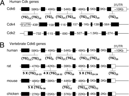
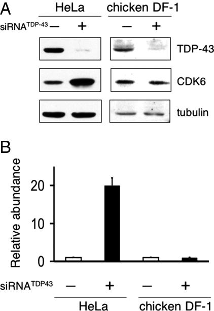
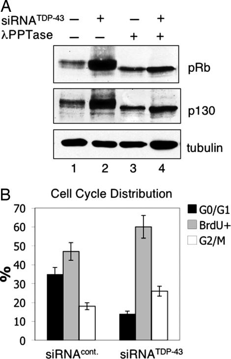
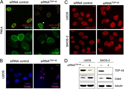
TDP-43 (for TAR DNA binding protein) is a highly conserved heterogeneous nuclear ribonucleoprotein (hnRNP) involved in specific pre-mRNA splicing and transcription events. TDP-43 recently has been identified as the main component of cytoplasmic inclusions in frontotemporal lobar degeneration (FTLD) and amyotrophic lateral sclerosis (ALS), two neurodegenerative disorders. The cellular role of this protein remains to be identified. Here, we show that loss of TDP-43 results in dysmorphic nuclear shape, misregulation of the cell cycle, and apoptosis. Removal of TDP-43 in human cells significantly increases cyclin-dependent kinase 6 (Cdk6) protein and transcript levels. The control of Cdk6 expression mediated by TDP-43 involves GT repeats in the target gene sequence. Cdk6 up-regulation in TDP-43-depleted cells is accompanied by an increase in phosphorylation of two of its major targets, the retinoblastoma protein pRb and pRb-related protein pRb2/p130. TDP-43 silencing also is followed by changes in the expression levels of several factors that control cell proliferation. Morphological nuclear defects and increased apoptosis upon TDP-43 loss are mediated via the pRb pathway because pRb-negative cells (Saos-2) do not undergo programmed cell death or nuclear shape deformation upon TDP-43 removal. Our results identify a regulatory target of TDP-43 and show that TDP-43 depletion has important consequences in essential metabolic processes in human cells.


Conflict of interest statement

The authors declare no conflict of interest.

Figures

Fig. 1.
Fig. 1.
GT repeats are unique to Cdk6 and are conserved in different mammals. Schematic representation of different Cdk genes, including coding exons (■) UTRs (□) and introns. (A) Human Cdk6 and the closely related Cdk4 and Cdk2 are shown. Highlighted in human Cdk6 are the GT repeat sequences. Two stretches ranging from 10 to 40 repeats are found in introns 1, 3, and 4. The repeats located at the 3′ UTR of the gene are 400 nt downstream of the stop codon and 180 nt upstream of the first polyadenylation site. In contrast to Cdk6, Cdk4, and Cdk2 have significantly shorter introns and 3′ UTR and lack GT repeat sequences. (B) TG repeats are present in primates, mouse, and rat Cdk6 genes. The length of the introns is more or less conserved, i.e., introns 1, 2, 3, and 4 are >35-Kb long, whereas introns 5 and 6 range from 1 to 5 Kb. Although chicken Cdk6 gene structure is similar to that of other vertebrates analyzed, the sequence lacks GT repeats entirely.
Fig. 2.
Fig. 2.
TDP-43 depletion causes up-regulation of Cdk6 in human but not in chicken cells. (A) Immunoblot analysis of HeLa and chicken embryo fibroblast (DF-1) cell extracts. Human and chicken cells were transfected with control or human/chicken TDP-43-targeted siRNA to check for changes in Cdk6 expression levels. Equal loading was confirmed by tubulin detection. Other human cell lines tested showed a similar increase in Cdk6 upon TDP-43 depletion. (B) Real-time RT-PCR analysis of Cdk6 cDNA in TDP-43 depleted (■) and control-treated (□) HeLa and DF-1 cells. Values shown are the average of three independent experiments. Error bars indicate SD.
Fig. 3.
Fig. 3.
TDP-43 depletion promotes pRb and p130 phosphorylation and causes changes in cell cycle distribution. (A) Western blot analysis to detect pRb and pRb2/p130 levels in U2OS cell extracts from TDP-43 depleted (lanes 2 and 4) and control treated cells (lanes 1 and 3). Extracts were also treated with λ phosphatase (PPTase) (lanes 3 and 4) to compare unphosphorylated pRb and pRb2/p130 protein levels. Tubulin was used as loading control. (B) Bars show the percentage of cells in G0/G1, S, and G2/M phases of the cell cycle in proliferating siRNATDP-43 and siRNA control-treated U2OS cells. Propidium- and BrdU-labeled cells were analyzed by FACS after RNAi treatment. Results show average values of three independent experiments, and error bars show SD.
Fig. 4.
Fig. 4.
Loss of TDP-43 results in nuclear membrane deformation and apoptosis. Immunofluorescence microscopy of siRNATDP-43 compared with control-treated cells is shown. (Scale bar, 10 μm.) (A) HeLa cells were stained with antibodies against lamin A/C and TDP-43 (red and green, Upper). Arrowhead points to a cell that escaped siRNA silencing. (Lower) Emerin-labeled nuclear membrane. Similar nuclear shape deformation was observed upon detection of lamin B and Lap2β. (B) U2OS cells were assayed for DAPI and TUNEL staining to detect apoptosis. (Scale bar, 10 μm.) (C) The nuclei of U2OS and Saos-2 cells were visualized by lamin A/C detection after TDP-43 depletion. (D) Down-regulation of TDP-43 in U2OS and Saos-2 cells and the consequent Cdk6 increase were verified by immunoblotting. Tubulin was used as loading control.

References

    1. Buratti E, Baralle FE. Characterization and functional implications of the RNA binding properties of nuclear factor TDP-43, a novel splicing regulator of CFTR exon 9. J Biol Chem. 2001;276:36337–36343. - PubMed
    1. Buratti E, Dork T, Zuccato E, Pagani F, Romano M, Baralle FE. Nuclear factor TDP-43 and SR proteins promote in vitro and in vivo CFTR exon 9 skipping. EMBO J. 2001;20:1774–1784. - PMC - PubMed
    1. Ayala YM, Pagani F, Baralle FE. TDP43 depletion rescues aberrant CFTR exon 9 skipping. FEBS Lett. 2006;580:1339–1344. - PubMed
    1. Buratti E, Brindisi A, Pagani F, Baralle FE. Nuclear factor TDP-43 binds to the polymorphic TG repeats in CFTR intron 8 and causes skipping of exon 9: A functional link with disease penetrance. Am J Hum Genet. 2004;74:1322–1325. - PMC - PubMed
    1. Mercado PA, Ayala YM, Romano M, Buratti E, Baralle FE. Depletion of TDP 43 overrides the need for exonic and intronic splicing enhancers in the human apoA-II gene. Nucleic Acids Res. 2005;33:6000–6010. - PMC - PubMed

Publication types

MeSH terms