. 2008 Mar 4;105(9):3256-61.
doi: 10.1073/pnas.0800254105. Epub 2008 Feb 27.

Phenylalanine-508 mediates a cytoplasmic-membrane domain contact in the CFTR 3D structure crucial to assembly and channel function

Affiliations

Phenylalanine-508 mediates a cytoplasmic-membrane domain contact in the CFTR 3D structure crucial to assembly and channel function

Adrian W R Serohijos et al. Proc Natl Acad Sci U S A. .

Abstract

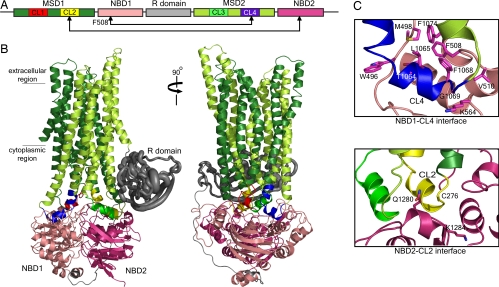
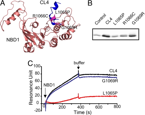
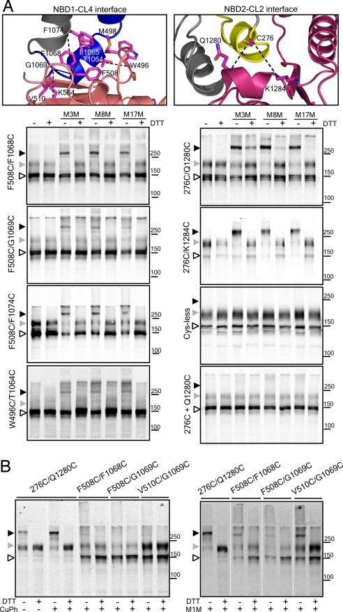
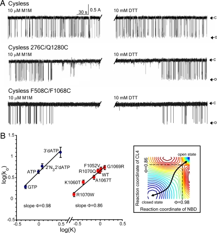
Deletion of phenylalanine-508 (Phe-508) from the N-terminal nucleotide-binding domain (NBD1) of the cystic fibrosis transmembrane conductance regulator (CFTR), a member of the ATP-binding cassette (ABC) transporter family, disrupts both its folding and function and causes most cystic fibrosis. Most mutant nascent chains do not pass quality control in the ER, and those that do remain thermally unstable, only partially functional, and are rapidly endocytosed and degraded. Although the lack of the Phe-508 peptide backbone diminishes the NBD1 folding yield, the absence of the aromatic side chain is primarily responsible for defective CFTR assembly and channel gating. However, the site of interdomain contact by the side chain is unknown as is the high-resolution 3D structure of the complete protein. Here we present a 3D structure of CFTR, constructed by molecular modeling and supported biochemically, in which Phe-508 mediates a tertiary interaction between the surface of NBD1 and a cytoplasmic loop (CL4) in the C-terminal membrane-spanning domain (MSD2). This crucial cytoplasmic membrane interface, which is dynamically involved in regulation of channel gating, explains the known sensitivity of CFTR assembly to many disease-associated mutations in CL4 as well as NBD1 and provides a sharply focused target for small molecules to treat CF. In addition to identifying a key intramolecular site to be repaired therapeutically, our findings advance understanding of CFTR structure and function and provide a platform for focused biochemical studies of other features of this unique ABC ion channel.


Conflict of interest statement

The authors declare no conflict of interest.

Figures

Fig. 1.
Fig. 1.
Theoretical model of CFTR structure. (A) Schema of CFTR primary structure containing two nucleotide-binding domains (NBD1 and NBD2), two membrane-spanning domains (MSD1 and MSD2), and a regulatory region (R domain). Each MSD contains two cytoplasmic loops (CL) that form interfaces with the NBDs. (B) Homology model of CFTR constructed from Sav1866 exporter (16) (see Results), where the domains are colored as in the schema. The unique-to-CFTR R domain, which is largely unstructured (23), was approximated by constructing an ensemble of dynamically accessible conformations derived from ab initio folding (see SI Text). R domain backbone size is rendered in proportion to variations of Cα atoms. (C) Close-up view of the interfaces formed between NBD1/CL4 and NBD2/CL2. Cross-linking of Cys pairs F508C/L1065C, F508C/F1068C, F508C/G1069C, and F508C/F1074C confirms that Phe-508 in NBD1 associates with CL4 in MSD2 (Fig. 3 and SI Fig. 7). Cross-linking of C276/Q1280C and C276/K1284C confirms interaction of CL2 and NBD2.
Fig. 2.
Fig. 2.
CL4 peptide binding to NBD1. (A) Location of disease-associated mutations (L1065P, R1066C, and G1069R) at the NBD1/CL4 interface. (B) Disease-causing mutations in CL4 abolish or diminish the CL4 and NBD1 interaction. Biotinylated CL4 or its mutant peptides immobilized on NeutrAvidin beads were incubated with purified recombinant human NBD1 (see SI Text). Bound proteins were eluted with sample buffer and detected by Western blotting with CFTR antibody 660. NeutrAvidin beads without bound peptide were used as control. (C) CL4 binds to NBD1 as detected with surface plasmon resonance. Biotinylated peptides were immobilized on a BIAcore streptavidin sensor chip to 200 resonance units. NBD1 was injected, and the binding was detected by surface plasmon resonance and BIAcore 2000. The binding of NBD1 to the chip without peptide was subtracted from NBD1 binding to the peptides.
Fig. 3.
Fig. 3.
Cross-linking of interfacial cysteine pairs. (A) Confirmation of the interaction, in cells, between NBD1 and CL4 (MSD2) and between NBD2 and CL2 (MSD1). Phe-508 participates in an apparent aromatic cluster with residues from CL4 (see also SI Fig. 10). CL4 also interacts with other regions in NBD1 as suggested by cross-linking of residues close to the Q loop (W496C/T1064C and M498C/L1065C) and a residue near the Walker B motif (K564C/G1069C). HEK293 cells transiently transfected with Cys-less CFTR containing Cys pairs were harvested and incubated in the presence of 200 μM MTS cross-linkers of different spacer arm lengths for 15 min at room temperature. Samples with or without DTT were subjected to SDS/PAGE and Western blotting with mAb596. Cross-linked species migrate above 250 kDa. (Right, Bottom two) Controls showing the lack of effects of cross-linking of the Cys-less construct and individually expressed and mixed single cysteine constructs (labeled 276C + Q1280C) under the same conditions (see also SI Fig. 8). (B) (Right) Cross-linking by the shortest reagent 1,1-methanediyl bismethanethiosulfonate (M1M), in isolated membranes, indicates close contact and probable mobility of residues across both interfaces. (Left) There is greater propensity for disulfide bond formation at the CL2/NBD2 interface (276C/Q1280C) than at the CL4/NBD1 interface. Open arrowhead, immature core-glycosylated CFTR (band B); gray arrowhead, mature complex-glycosylated CFTR (band C); black arrowhead, cross-linked mature protein.
Fig. 4.
Fig. 4.
Role of domain–domain interactions in CFTR channel gating. (A) Inhibition of CFTR channel gating by cross-linking. Single-channel recording after exposure to 10 μM M1M from the cis side of the bilayer followed by 10 mM DTT. For Cys-less CFTR, the last 4 min from the total 20 min of M1M treatment is shown in the first half of the trace before the interruption. The last 4 min of the total 20 min of DTT treatment is shown in the second half. O, open state; c, closed state. (Top) Cys-less CFTR (n = 4). (Middle) Cys-less CFTR with 276C/Q1280C (n = 3). (Bottom) Cys-less CFTR with F508C/F1068C (n = 4). (Middle and Bottom) Change in functional state in the middle of a 4-min portion of the total 20-min recording. No effect of MTS reagents was observed in constructs containing single cysteines that contribute to cross-linkable pairs (SI Fig. 11). (B) NBD1 and CL4 participate early in the gating cycle. Brønsted plots for wild-type CFTR gated by 2 mM nucleotide ligands shown by each experimental point (Left) and substitutions of CL4 residues (Right). Both graphs are linear with the slope Φ values indicated. Points on both graphs are shown as mean values ± SEM of at least four different experiments. (Inset) Hypothetical free energy landscape of CFTR gating where troughs (stable states) are colored blue and crests (unstable states) are red. At the transition state (saddle point), NBD1 has proceeded 98% toward the open state, and CL4 has proceeded 86%.

References

    1. Quinton PM. Cystic fibrosis: Lessons from the sweat gland. Physiology. 2007;22:212–225. - PubMed
    1. Al Awqati Q. Alternative treatment for secretory diarrhea revealed in a new class of CFTR inhibitors. J Clin Invest. 2002;110:1599–1601. - PMC - PubMed
    1. Davis PB. Cystic fibrosis since 1938. Am J Respir Crit Care Med. 2006;173:475–482. - PubMed
    1. Bear CE, et al. Purification and functional reconstitution of the cystic fibrosis transmembrane conductance regulator (CFTR). Cell. 1992;68:809–818. - PubMed
    1. Gadsby DC, Vergani P, Csanady L. The ABC protein turned chloride channel whose failure causes cystic fibrosis. Nature. 2006;440:477–483. - PMC - PubMed

Publication types

MeSH terms

Substances

LinkOut - more resources