. 2008 Mar 4;105(9):3327-32.
doi: 10.1073/pnas.0711864105. Epub 2008 Feb 27.

Yeast Pch2 promotes domainal axis organization, timely recombination progression, and arrest of defective recombinosomes during meiosis

Affiliations

Yeast Pch2 promotes domainal axis organization, timely recombination progression, and arrest of defective recombinosomes during meiosis

G Valentin Börner et al. Proc Natl Acad Sci U S A. .

Abstract

We show that, during budding yeast meiosis, axis ensemble Hop1/Red1 and synaptonemal complex (SC) component Zip1 tend to occur in alternating strongly staining domains. The widely conserved AAA+-ATPase Pch2 mediates this pattern, likely by means of direct intervention along axes. Pch2 also coordinately promotes timely progression of cross-over (CO) and noncross-over (NCO) recombination. Oppositely, in a checkpoint-triggering aberrant situation (zip1Delta), Pch2 mediates robust arrest of stalled recombination complexes, likely via nucleolar localization. We suggest that, during WT meiosis, Pch2 promotes progression of SC-associated CO and NCO recombination complexes at a regulated early-midpachytene transition that is rate-limiting for later events; in contrast, during defective meiosis, Pch2 ensures that aberrant recombination complexes fail to progress so that intermediates can be harmlessly repaired during eventual return to growth. Positive vs. negative roles of Pch2 in the two situations are analogous to positive vs. negative roles of Mec1/ATR, suggesting that Pch2 might mediate Mec1/ATR activity. We further propose that regulatory surveillance of normal and abnormal interchromosomal interactions in mitotic and meiotic cells may involve "structure-dependent interchromosomal interaction" (SDIX) checkpoints.


Conflict of interest statement

The authors declare no conflict of interest.

Figures

Fig. 1.
Fig. 1.
Localization of Zip1, Hop1, and Red1 along meiotic chromosomes in WT and pch2Δ meiosis. (A) Zip1-GFP localization in WT (SEY674). Filled and open arrowheads indicate regions of greater or lesser abundance, respectively. (B) Hop1 and Red1 localization in WT (NKY3330). (C) Zip1 and Hop1 staining in WT and pch2Δ. Pachytene in WT (a–d) and pch2Δ (e–h); zygotene in WT (i–k), and pch2Δ (m–o); leptotene in WT (l) and pch2Δ (p). WT (VBY338), pch2Δ (VBY1026). (D) Degree of overlap between strong Hop1 and Zip1 signals during zygotene and pachytene in WT (Left) and pch2Δ (Right). Zygotene (WT, t = 5, 6, and 7 h; n = 21, 12, and 11, respectively; pch2Δ, t = 5, 6, and 7 h; n = 26, 17, and 23, respectively) pachytene (WT, t = 5, 6, 7, and 8.5 h; n = 11, 18, 11, and 4, respectively; pch2Δ, t = 5, 6, 7, and 8.5 h; n = 6, 16, 12, and 25, respectively). (E) Abundance of continuous Hop1 lines in WT (black) and pch2Δ (red).
Fig. 2.
Fig. 2.
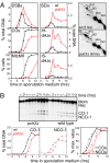
Recombination at the HIS4LEU2 recombination hotspot in WT and pch2Δ. (A) (Left) Quantitative analysis of recombination intermediates and products. DSBs and COs were quantitated from 1D gels; SEIs, interhomolog (IH)-dHJs, and intersister (IS)-dHJs were quantitated from 2D gels. (Right) Representative time points from 2D gels. # indicates long joint molecules (26) (see SI Fig. 8 C and D for complete gel images). Cells that had completed meiosis I (MI±MII) were identified by determining the number of cells with two to four DAPI-staining nuclei. WT (VBY310), pch2Δ (VBY311). (B) (Upper) Southern blot analysis of a 1D gel after digestion with XhoI and MluI. (Lower) Quantification of blot shown in Upper. (Lower Left) Absolute amounts of CO-1 and NCO-1 recombination products (SI Fig. 8A). (Lower Right) The same data plotted as a percentage of maximum levels to compare kinetics of CO and NCO formation. WT (VBY338); pch2Δ (VBY1026).
Fig. 3.
Fig. 3.
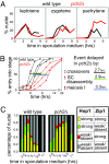
Pachytene exit in WT and pch2Δ. (A) Kinetics of Zip1 loading and unloading in WT (black) and pch2Δ (red). More than 150 nuclei per time point were classified as leptotene, zygotene, and pachytene as shown in Fig. 1C a, i, and l. Leptotene and zygotene occur in pch2Δ with kinetics similar to WT. Nuclei with lines of Zip1 appear and disappear in a timely manner in the WT, yet accumulate and persist in pch2Δ at high levels; WT (VBY338), pch2Δ (VBY1026). (B) Relative timing of crossing over, pachytene exit, and first meiotic division. All data are from parallel WT and pch2Δ time courses, respectively (time course 84). Cross-overs and MI±MII are plotted as percentage of maximum levels. The cumulative curve for pachytene exit was calculated by processing primary pachytene curves from Fig. 3A as described in ref. . Ninety percent of cells were assumed to enter pachytene in both WT and pch2Δ, and pachytene was assumed to end at 24 h in pch2Δ, consistent with absence of nuclei containing Zip1 staining in pch2Δ at t = 24 h. (C) Zip1 and Hop1 loading kinetics in WT and pch2Δ. Nuclei (n ≥ 30) were classified in surface-spread nuclei immunodecorated with appropriate antibodies at the indicated time points based on levels of Hop1 and Zip1 staining. Nuclei with strong Hop1 and Zip1 loading (yellow) start decreasing in the WT at 6 h but accumulate and persist in pch2Δ. Nuclei with only Zip1 or Hop1 are rare in the WT at late time points, yet a class carrying only strong Hop1 staining (green) reappears at late time points in pch2Δ, suggesting delayed unloading of Hop1.
Fig. 4.
Fig. 4.
Effect of pch2Δ on recombination and meiotic progression in zip1Δ. (A) Meiotic divisions are partially restored in pch2Δ, with half of the cells progressing through meiosis with an ≈3-h delay, and the other half still exhibiting arrest (Left). Cross-over formation is partially restored in the zip1Δpch2Δ mutant (Right). (B) Quantitation of DSBs, SEIs, IH-dHJs, and IS-dHJs in WT, zip1Δ, and zip1Δpch2Δ (time course #90). WT (VBY310), zip1Δ (VBY1050), zip1Δpch2Δ (VBY312). (C) Ratios of IS/IH-dHJs in time course #90. See also SI Fig. 9.
Fig. 5.
Fig. 5.
Absence of Sir2 results in a pch2Δ-like mutant phenotype only in a zip1Δ mutant background. (A) (Left) Formation of CO products (solid lines) and meiotic progression (dashed lines) in WT (black) and sir2Δ (green). COs were determined at the HIS4LEU2 hotspot and quantitated from a 1D gel. (Right) Levels of DSBs are from a 1D gel; SEI, IH-dHJ, and IS-dHJs were determined from 2D gels. Increased IH-dHJ levels in sir2Δ are detected only at a single time point and are likely due to an associated increase in CO products. Data are from time course #71. WT (VBY310), sir2Δ (VBY945). (B) Hop1 and Zip1 localization in sir2Δ pachytene nuclei. See Fig. 1C a–h for images of pachytene images and classes. For sir2Δ, pachytene nuclei (n = 14) at t = 6 h were analyzed for colocalization of strong Hop1 and Zip1 signals (data for WT and pch2Δ from Fig. 1D). Pachytene nuclei at t = 6 h are observed at the following frequencies: 14% (WT), 22% (sir2Δ), 40% (pch2Δ). Hop1 lines per class d at t = 6 h are observed with the following frequencies: 4% (WT), <1% (sir2Δ), and 31% (pch2Δ; n > 150 for WT and mutants). (C) Meiotic progression and recombination in zip1Δ and zip1Δsir2Δ. (D) Ratios of IS/IH-dHJs in zip1Δsir2Δ and zip1Δ (time course #90).

References

    1. Sym M, Engebrecht JA, Roeder GS. ZIP1 is a synaptonemal complex protein required for meiotic chromosome synapsis. Cell. 1993;72:365–378. - PubMed
    1. Smith AV, Roeder GS. The yeast Red1 protein localizes to the cores of meiotic chromosomes. J Cell Biol. 1997;136:957–967. - PMC - PubMed
    1. Blat Y, Protacio RU, Hunter N, Kleckner N. Physical and functional interactions among basic chromosome organizational features govern early steps of meiotic chiasma formation. Cell. 2002;111:791–802. - PubMed
    1. Keeney S. Mechanism and control of meiotic recombination initiation. Curr Top Dev Biol. 2001;52:1–53. - PubMed
    1. Tesse S, Storlazzi A, Kleckner N, Gargano S, Zickler D. Localization and roles of Ski8p protein in Sordaria meiosis and delineation of three mechanistically distinct steps of meiotic homolog juxtaposition. Proc Natl Acad Sci USA. 2003;100:12865–12870. - PMC - PubMed

Publication types

MeSH terms