. 2008;47(13):2394-7.
doi: 10.1002/anie.200704847.

A FRET-based fluorogenic phosphine for live-cell imaging with the Staudinger ligation

Affiliations

A FRET-based fluorogenic phosphine for live-cell imaging with the Staudinger ligation

Matthew J Hangauer et al. Angew Chem Int Ed Engl. 2008.
No abstract available


Figures

Figure 1
Figure 1
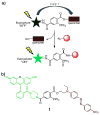
A FRET-based fluorogenic phosphine for live cell imaging. a) The design of a quenched phosphine-fluorophore that is activated upon Staudinger ligation with azides. b) Compound 1, possessing fluorescein (green) and disperse red 1 (dark red) moieties.
Figure 2
Figure 2
Labeling of azido-mDHFR with compound 1. Purified azido-mDHFR (left lane) and native mDHFR (right lane) were labeled with 12.5 μM 1 for 20 hours at RT in PBS under denaturing conditions. The crude reaction mixtures were separated by SDS-PAGE and the gel was analyzed by fluorescence imaging (top row) and by Zn stain to reveal total protein content (bottom row).
Figure 3
Figure 3
Flow cytometry analysis of live Ac4ManNAz-treated CHO cells labeled by phosphine 1. Cells were treated with Ac4ManNAz for 3 days and then phosphine 1 for 8 hours. Control cells were either untreated, or treated with phosphine 1 alone. Error bars represent the standard deviation of the mean for triplicate measurements.
Figure 4
Figure 4
Fluorescence microscopy of Ac4ManNAz-treated HeLa cells labeled with phosphine 1. a) Top row: live HeLa cells treated with Ac4ManNAz, 1 (FITC channel), and the live cell Golgi marker BODIPY® TR C5-ceramide (Cy3 channel); bottom row: HeLa cells treated with Ac4ManNAz, 1, then fixed, permeabilized and treated with anti-Golgin 97 mouse mAb and goat anti-mouse IgG-Alexa Fluor 647 (Cy5 channel). b) Live HeLa cells treated with 1 only. All cells were treated while alive with nuclear stain Hoechst 33342 (DAPI channel) and viability stain propidium iodide (Cy3 and Cy5 channels). The lack of nuclear fluorescence in Cy3 and Cy5 channels indicates propidium iodide exclusion from cells. The scale bars represent 10 μm.
Scheme 1
Scheme 1
Synthesis of phosphine 1. Reagents and conditions: a) tBuOH, DMAP (0.5 equiv), EDAC, CH2Cl2 (84%); b) LiOH (1.5 equiv), 3:1 MeOH:H2O (94%); c) HPPh2, K2CO3, Pd(OAc)2 (0.3 mol %), CH3CN, reflux (72%); d) disperse red 1, DMAP (0.1 equiv), DCC, CH3CN (83%); e) TFA (26 equiv), TES (5 equiv), CH2Cl2 (87%); f) 8, HATU, DIPEA, DMF (65%).
Scheme 2
Scheme 2
Model Staudinger ligation.

References

    1. O’Hare HM, Johnsson K, Gautier A. Curr Opin Struct Biol. 2007;17:488. - PubMed
    2. Straight AF. Methods Cell Biol. 2007;81:93. - PubMed
    1. Ohtsubo K, Marth JD. Cell. 2006;126:855. - PubMed
    2. Casey PJ. Science. 1995;268:221. - PubMed
    1. Prescher JA, Bertozzi CR. Nat Chem Biol. 2005;1:13. - PubMed
    1. Link AJ, Vink MK, Tirrell DA. J Am Chem Soc. 2004;126:10598. - PubMed
    2. Kirshenbaum K, Carrico IS, Tirrell DA. Chembiochem. 2002;3:235. - PubMed
    3. Chin JW, Santoro SW, Martin AB, King DS, Wang L, Schultz PG. J Am Chem Soc. 2002;124:9026. - PubMed
    1. Laughlin ST, et al. Methods Enzymol. 2006;415:230., see Supporting Information; Rabuka D, Hubbard SC, Laughlin ST, Argade SP, Bertozzi CR. J Am Chem Soc. 2006;128:12078.Sawa M, Hsu TL, Itoh T, Sugiyama M, Hanson SR, Vogt PK, Wong CH. Proc Natl Acad Sci U S A. 2006;103:12371.

Publication types

MeSH terms

LinkOut - more resources