

Orthotopic implantation mouse model and cDNA microarray analysis indicates several genes potentially involved in lymph node metastasis of colorectal cancer
- PMID: 18307535
- PMCID: PMC11158708
- DOI: 10.1111/j.1349-7006.2008.00725.x
Orthotopic implantation mouse model and cDNA microarray analysis indicates several genes potentially involved in lymph node metastasis of colorectal cancer
Abstract
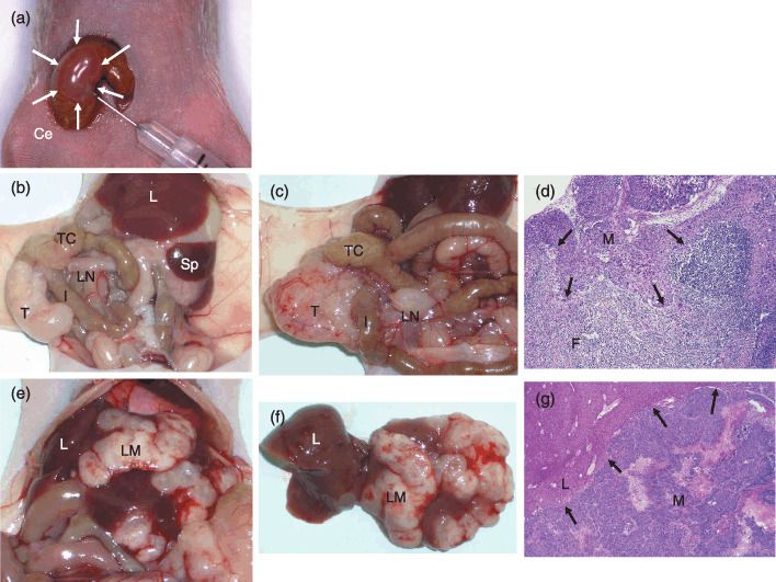
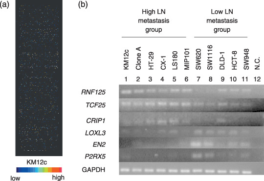
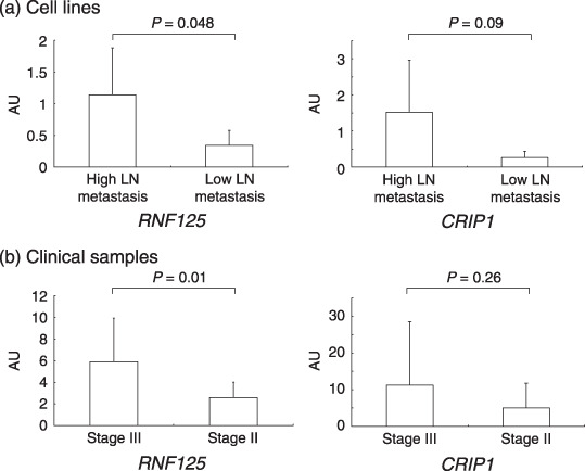
In colorectal cancer (CRC) patients, metastasis to the regional lymph node (LN) is an important first step in the dissemination of cancers. To identify the genes possibly involved in LN metastasis of CRC, we analyzed LN metastases in an orthotopic implantation mouse model with 22 CRC cell lines using Matrigel, an extracellular matrix protein derived from mice sarcoma, and combined the data with gene expression profiles of cDNA microarray of those cell lines. With this implantation analysis, the incidence of LN metastasis was 60% in 228 orthotopically implanted mice and varied from 100% to 0% among the cell lines. KM12c and Clone A showed LN metastasis in all orthotopically implanted mice, but DLD-1, HCT-8, and SW948 did not show LN metastases at all. In contrast, the incidence of liver and lung metastasis in 22 CRC cell lines was 13% and 1%, respectively. Combining those data with cDNA microarray in vitro, we isolated 636 genes that were differentially expressed depending on the incidence of LN metastasis. Among those genes, the expression level of ring finger protein 125 (RNF125), previously known as an E3 ubiquitin ligase in T cell activation, was significantly different between primary tumors in Stage III CRC patients with LN metastasis and Stage II patients without LN metastasis. In conclusion, the orthotopic implantation mice model with Matrigel was useful, and we isolated candidate genes such as RNF125 that possibly play an important role in LN metastasis of CRC cells.
Figures
Similar articles
-
Differential metastasis-associated gene analysis of prostate carcinoma cells derived from primary tumor and spontaneous lymphatic metastasis in nude mice with orthotopic implantation of PC-3M cells.Cancer Lett. 2006 Feb 20;233(1):79-88. doi: 10.1016/j.canlet.2005.03.034. Cancer Lett. 2006. PMID: 15885894
-
Down-regulated expression of SATB2 is associated with metastasis and poor prognosis in colorectal cancer.J Pathol. 2009 Sep;219(1):114-22. doi: 10.1002/path.2575. J Pathol. 2009. PMID: 19557828
-
Gene expression profiling in lymph node-positive and lymph node-negative colorectal cancer.Dis Colon Rectum. 2004 Feb;47(2):141-52. doi: 10.1007/s10350-003-0032-7. Dis Colon Rectum. 2004. PMID: 15043283
-
Restoration of PTEN activity decreases metastases in an orthotopic model of colon cancer.J Surg Res. 2013 Oct;184(2):755-60. doi: 10.1016/j.jss.2013.03.035. Epub 2013 Apr 6. J Surg Res. 2013. PMID: 23623571 Free PMC article.
-
Microarray analysis reveals that high mobility group A1 is involved in colorectal cancer metastasis.Oncol Rep. 2013 Sep;30(3):1488-96. doi: 10.3892/or.2013.2602. Epub 2013 Jul 8. Oncol Rep. 2013. PMID: 23835740
Cited by
-
The oncogenic role of LncRNA FAM83C-AS1 in colorectal cancer development by epigenetically inhibits SEMA3F via stabilizing EZH2.Aging (Albany NY). 2020 Oct 27;12(20):20396-20412. doi: 10.18632/aging.103835. Epub 2020 Oct 27. Aging (Albany NY). 2020. PMID: 33109776 Free PMC article.
-
Downregulation of miR-193a-5p correlates with lymph node metastasis and poor prognosis in colorectal cancer.World J Gastroenterol. 2014 Sep 14;20(34):12241-8. doi: 10.3748/wjg.v20.i34.12241. World J Gastroenterol. 2014. PMID: 25232258 Free PMC article.
-
Activation of Slit2/Robo1 Signaling Promotes Tumor Metastasis in Colorectal Carcinoma through Activation of the TGF-β/Smads Pathway.Cells. 2019 Jun 25;8(6):635. doi: 10.3390/cells8060635. Cells. 2019. PMID: 31242633 Free PMC article.
-
Cytotoxic effect of different treatment parameters in pressurized intraperitoneal aerosol chemotherapy (PIPAC) on the in vitro proliferation of human colonic cancer cells.World J Surg Oncol. 2017 Feb 10;15(1):43. doi: 10.1186/s12957-017-1109-4. World J Surg Oncol. 2017. PMID: 28183319 Free PMC article.
-
Slug expression enhances tumor formation in a noninvasive rectal cancer model.J Surg Res. 2011 Sep;170(1):56-63. doi: 10.1016/j.jss.2011.02.012. Epub 2011 Mar 23. J Surg Res. 2011. PMID: 21470622 Free PMC article.
References
-
- Ajiki W, Tsukuma H, Oshima A. Cancer incidence and incidence rates in Japan in 1999: estimates based on data from 11 population‐based cancer registries. Jpn J Clin Oncol 2004; 34: 352–6. - PubMed
-
- Statistics and Information Department Minister's Secretariat, Ministry of Health Labour and Welfare . Vital Statistics in Japan, Trends Up to 2004. Tokyo, Japan: Health and Welfere Statistics Association Publisher, 2006. (in Japanese).
-
- Japanese Society for Cancer of the Colon and Rectum . Treatment Guidelines on Cancer of the Colon, Rectum and Anus. Tokyo, Japan: Kanehara Publishers, 2005. (in Japanese).
-
- Kleinman HK, McGarvey ML, Liotta LA, Robey PG, Tryggvason K, Martin GR. Isolation and characterization of type IV procollagen, laminin, and heparan sulfate proteoglycan from the EHS sarcoma. Biochemistry 1982; 21: 6188–93. - PubMed
-
- Yoshida Y, Kamitani N, Sasaki H, Kusumi K, Tominaga T, Kotsuji F. Establishment of a liver metastatic model of human ovarian cancer. Anticancer Res 1998; 18: 327–31. - PubMed
Publication types
MeSH terms
LinkOut - more resources
Full Text Sources
Medical
Research Materials
Miscellaneous
