. 2008 Apr;99(4):762-9.
doi: 10.1111/j.1349-7006.2008.00739.x. Epub 2008 Feb 27.

The human endonuclease III enzyme is a relevant target to potentiate cisplatin cytotoxicity in Y-box-binding protein-1 overexpressing tumor cells

Affiliations

The human endonuclease III enzyme is a relevant target to potentiate cisplatin cytotoxicity in Y-box-binding protein-1 overexpressing tumor cells

David Guay et al. Cancer Sci. 2008 Apr.

Abstract

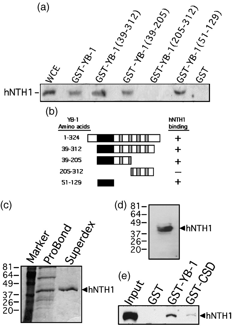
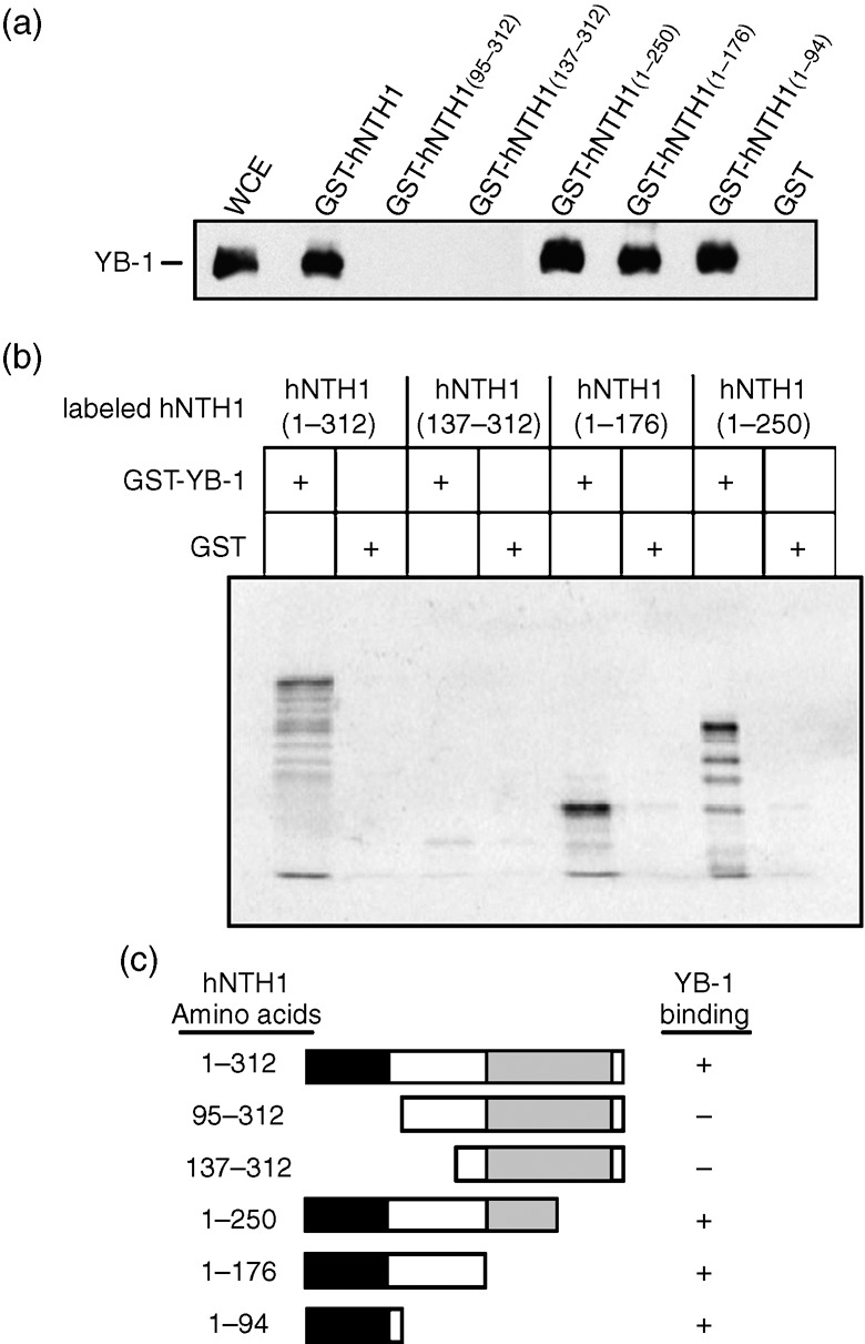
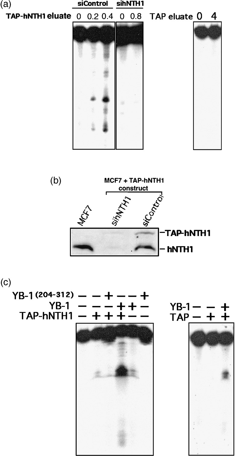
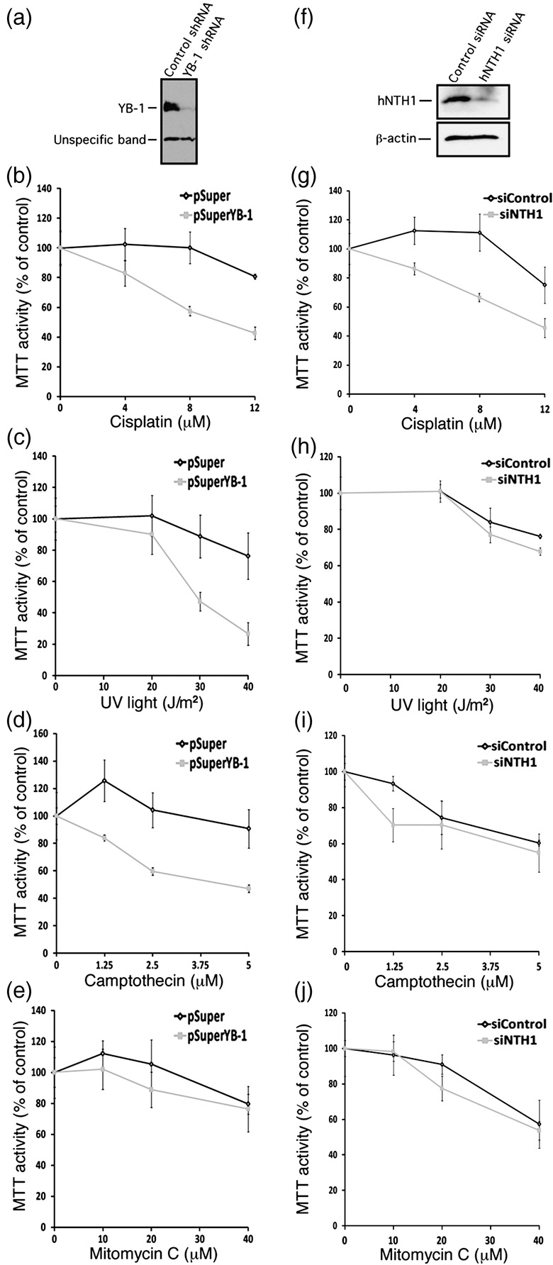
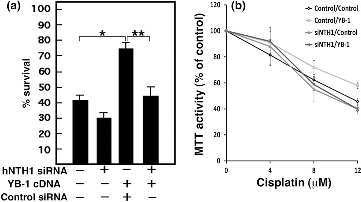
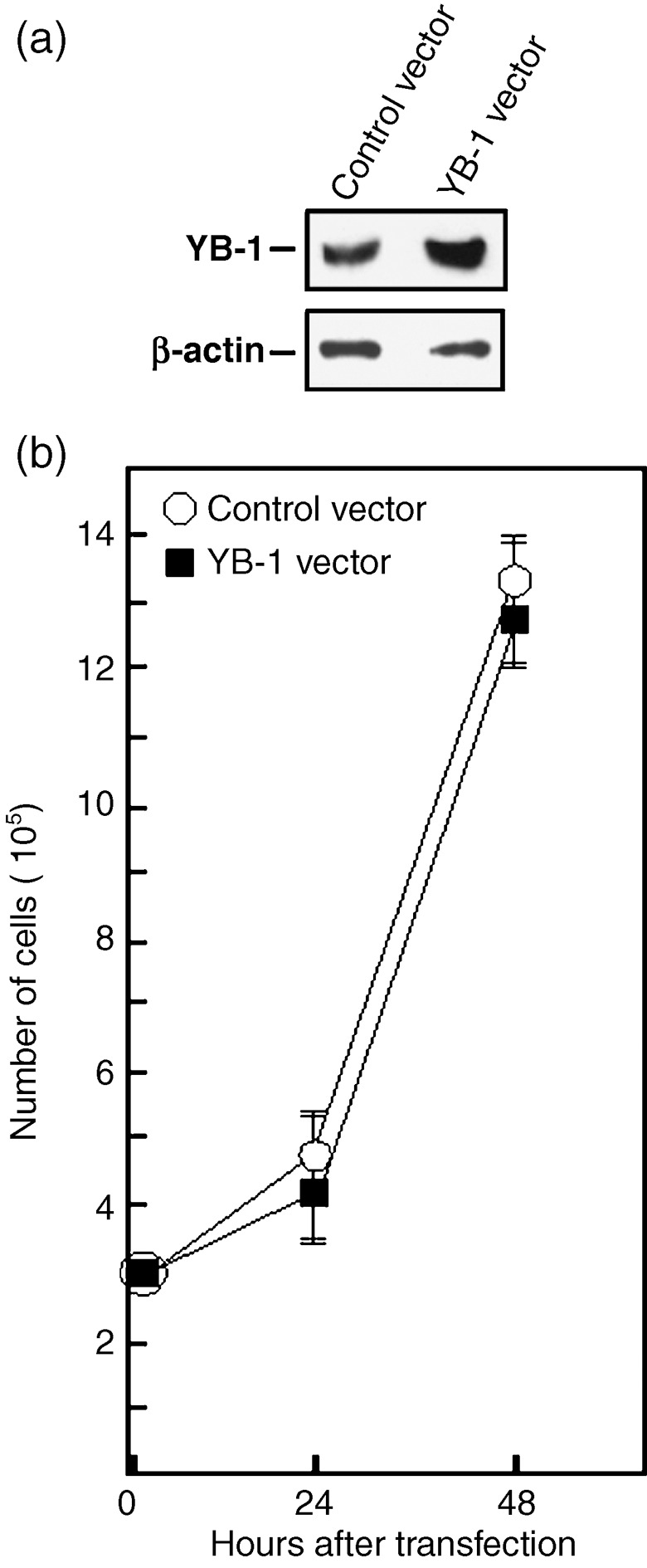
Y-box-binding protein-1 (YB-1) is a multifunctional protein involved in the regulation of transcription, translation, and mRNA splicing. In recent years, several laboratories have demonstrated that YB-1 is directly involved in the cellular response to genotoxic stress. Importantly, YB-1 is increased in tumor cell lines resistant to cisplatin, and the level of nuclear expression of YB-1 is predictive of drug resistance and patient outcome in breast tumors, ovarian cancers, and synovial sarcomas. YB-1 binds to several DNA repair enzymes in vitro including human endonuclease III (hNTH1). Human NTH1 is a bifunctional DNA glycosylase/apurinic/apyrimidinic lyase involved in base excision repair. In this study, we show that YB-1 binds specifically to the auto-inhibitory domain of hNTH1, providing a mechanism by which YB-1 stimulates hNTH1 activity. Indeed, YB-1 strongly stimulates in vitro the activity of hNTH1 toward DNA duplex probes containing oxidized bases, lesions prone to be present in cisplatin treated cells. We also observed an increase in YB-1/hNTH1 complex formation in the mammary adenocarcinoma MCF7 cell line treated with UV light and cisplatin. Such an increase was not observed with mitomycin C or the topoisomerase I inhibitor camptothecin. Accordingly, antisense RNAs against either YB-1 or hNTH1 increased cellular sensitivity to UV and cisplatin but not to mitomycin C. An antisense RNA against YB-1 increased camptothecin sensitivity. In contrast, an antisense against hNTH1 did not. Finally, siRNA against hNTH1 re-established cytotoxicity in otherwise cisplatin-resistant YB-1 overexpressing MCF7 cells. These data indicate that hNTH1 is a relevant target to potentiate cisplatin cytotoxicity in YB-1 overexpressing tumor cells.


Figures

Figure 1
Figure 1
Interaction of different domains of Y‐box‐binding protein‐1 (YB‐1) with human endonuclease III (hNTH1) in total cell extract. (a) Immunoblot against hNTH1 proteins bound to different GST‐YB‐1 affinity Sepahrose beads. Human MCF7 whole cell extracts (WCE) were incubated with either 50 µg of GST‐YB‐1 or GST‐linked glutathione‐sepharose beads overnight. Proteins bound to the affinity beads were analyzed by SDS‐PAGE with antibodies against hNTH1. (b) Schematic representation of different YB‐1 polypeptides that were used in the YB‐1 affinity chromatography experiments. The black box is the cold shock domain and the gray boxes are the basic/acidic cluster domains. The amino acid residues of the YB‐1 fragments used in this study are indicated on the left. hNTH1 binding is indicated on the right by the ‘+’ sign. The ‘–’ sign indicates no binding detected. (c) Coomassie staining of a gel containing purified hNTH1 after the nickel column step (ProBond resin) and after fractionation on Superdex‐200 (fraction number 29). The molecular weight, in kDa, is indicated on the left. (d) Western blot of purified hNTH1 after the final Superdex‐200 step (fraction 29) with an antibody against hNTH1. (e) Interaction of purified hNTH1 (after the final Superdex‐200 step) with 50 µg of GST‐YB‐1, GST‐CSD (cold shock domain of YB‐1; amino acids 51–129), or GST‐linked glutathione‐sepharose beads.
Figure 2
Figure 2
Interaction of the auto‐inhibitory domain of human endonuclease III (hNTH1) with the Y‐box‐binding protein‐1 (YB‐1) protein. (a) Immunoblot against YB‐1 proteins bound to different GST‐hNTH1 affinity Sepahrose beads. Human MCF7 cell extracts (WCE) were incubated with either 50 µg of GST‐hNTH1 constructs or GST‐linked glutathione‐sepharose beads overnight. Proteins bound to the affinity beads were analyzed by SDS‐PAGE with antibodies against YB‐1. (b) IVTT/GST interaction assay. In vitro‐transcribed/translated 35S‐labeled hNTH1 protein fragments bound to YB‐1‐GST affinity beads were separated by SDS‐PAGE and visualized using a PhosphoImager. (c) Schematic representation of the different hNTH1 polypeptides that were used in the hNTH1 affinity chromatography experiments. The black box is the auto‐inhibitory domain and the gray box is the catalytic domain. The amino acid residues of the hNTH1 fragments used in this study are indicated on the left. YB‐1 binding is indicated on the right by the ‘+’ sign. The ‘–’ sign indicates no binding detected.
Figure 3
Figure 3
Nuclease activity of eluted TAP‐hNTH1 in the presence of purified Y‐box‐binding protein‐1 (YB‐1). (a) Tap‐hNTH1 MCF7 expressing cells were transfected with either siRNA control (siControl) or siRNA against human endonuclease III (hNTH1) (panels on the left). Two days later the TAP‐hNTH1 complex was purified on streptavidin column and eluted with biotin. Eluted TAP‐hNTH1 proteins (0, 0.2, or 0.4 µg) were incubated for 30 min at 37°C as described in ‘Materials and Methods’ with a radioactive DNA duplex containing 8‐oxoguanine residues. Reactions were stopped in the appropriate dye buffer and cleaved DNA products were analyzed on 14% denaturing polyacrylamide gels. The panel on the right represents nuclease assays with or without eluate from MCF7 cells containing an empty TAP vector (0 or 0.8 µg). (b) Western blot showing intracellular levels of endogenous hNTH1 and TAP‐hNTH1 after transfection with a siRNA specific to hNTH1 or with a scrambled control siRNA. The first lane contained whole lysate from untransfected parental MCF7 cells. Each lane contains 75 µg of whole cell lysate. (c) Nuclease activity with TAP‐hNTH1 in the absence or presence of either a full length YB‐1 peptide (2 µg) or a purified peptide containing amino acids 205–312 of YB‐1 (2 µg). (Panel on the left.) Note that YB‐1(205–312) does not bind to hNTH1. The panel on the right represents nuclease assays with eluate from MCF7 cells containing an empty TAP vector (0.8 µg) in the presence or absence of YB‐1 (2 µg).
Figure 4
Figure 4
Co‐precipitation of human endonuclease III (hNTH1) protein with TAP‐YB‐1 protein under different DNA‐damaging conditions. (a) Detection of endogenous hNTH1 and Y‐box‐binding protein‐1 (YB‐1) proteins in whole cell lystes after transfection of TAP‐YB‐1 construct into MCF7 cells. (b) Detection of TAP‐YB‐1 and hNTH1 proteins after streptavidin affinity precipitation of the TAP constructs. (c–f) Detection of TAP‐YB‐1 and hNTH1 proteins after streptavidin affinity precipitation of the TAP constructs in untreated cells and in MCF7 cells treated (4 h) with either increasing doses of cisplatin (c), UV light (d), mitomycin C (e), or camptothecin (f). Under each Western blot, scanning analyses of the Western blots (from two independent experiments) are presented. Data are expressed as the mean (±SD) ratio of hNTH1 signals over the affinity‐precipitated TAP‐YB‐1 signals.
Figure 5
Figure 5
Impact of depleting either endogenous Y‐box‐binding protein‐1 (YB‐1) or human endonuclease III (hNTH1) proteins in MCF7 on survival after different DNA‐damaging conditions. (a) Western blots showing intracellular levels of YB‐1 48 h after transfection with a shRNA specific to YB‐1 or with a control construct. The lower molecular weight band is an unspecific protein recognized by the anti‐YB‐1 antibody. (b–e) MTT cell survival assays of MCF7 cells transfected with a shRNA specific to YB‐1 or with a control construct after of treatment with either cisplatin (b), UV light (c), camptothecin (d), or mitomycin C (e). Cells were first transfected with the appropriate constructs. Forty‐eight hours later, cells were plated in 96‐well plates. Cells were then treated with the drugs for 16 h in culture before the MTT assays. Results represent the mean ± SD of triplicate experiments. (f) Western blots showing intracellular levels of hNTH1 after 48 h after transfection with a siRNA specific to hNTH1 or with a scrambled control siRNA. The anti β‐actin was used as loading control for the blot. (g–j) MTT cell survival assays of MCF7 cells transfected with a siRNA specific to hNTH1 or with a scrambled control siRNA after treatment with either (g) cisplatin, (h) UV light, (i) camptothecin, or (j) mitomycin C. Cells were first transfected with the siRNAs. Forty‐eight hours later, cells were plated in 96‐well plates. Cells were then treated with the drugs for 16 h in culture before the MTT assays. Results represent the mean ± SD of triplicate experiments.
Figure 6
Figure 6
Impact of depleting endogenous human endonuclease III (hNTH1) proteins in Y‐box‐binding protein‐1 (YB‐1) overexpressing MCF7 cisplatin‐resistant cells. MCF7 cells were transfected with a siRNA specific to hNTH1 or scrambled control siRNA. Forty‐eight hours later, cells were transfected with YB‐1 expression vector or empty expression control vector. (a) Transfected cells were treated with 4 µM cisplatin for 16 h before counting with a hemacytometer (*P < 0.02; P < 0.05). The ‘–’ sign indicates scrambled siRNA or empty expression vector. The ‘+’ sign indicates sihNTH1 or YB‐1 expression vector. Experiments were done in triplicates. (b) Transfected cells were plated in 96‐well plates. Cells were then treated with the drugs for 16 h in culture before the MTT assays. Results represent the mean ± SD of the triplicate experiments.
Figure 7
Figure 7
Cell growth of MCF7 cells overexpressing Y‐box‐binding protein‐1 (YB‐1). (a) Example of Western blots showing intracellular levels of YB‐1 48 h after transfection with either an empty expression vector or a YB‐1 expression vector. The anti β‐actin is used as loading control for the blot (bottom panel). On average, transfection of YB‐1 expression vector increased the levels of YB‐1 proteins by two‐fold. (b) Cell growth of transfected MCF7 cells. Cells were transfected with either an empty expression vector or a YB‐1 expression vector by electroporation with a nucleofector kit (see ‘Materials and Methods’ for details). Approximately, 3 × 105 transfected cells were plated on 90‐mm Petri dishes. Cells were counted with an hemocytometer 24 and 48 h after the transfections. Results represent the mean ± SD of four independent transfection experiments. All cultures reached confluence at 72 h (data not shown).

Similar articles

Cited by

References

    1. Swamynathan SK, Nambiar A, Guntaka RV. Role of single‐stranded DNA regions and Y‐box proteins in transcriptional regulation of viral and cellular genes. FASEB J 1998; 12: 515–22. - PubMed
    1. Ashizuka M, Fukuda T, Nakamura T et al . Novel translational control through an iron–responsive element by interaction of multifunctional protein YB‐1 and IRP2. Mol Cell Biol 2002; 22: 6375–83. - PMC - PubMed
    1. Stickeler E, Fraser SD, Honig A, Chen AL, Berget SM, Cooper TA. The RNA binding protein YB‐1 binds A/C‐rich exon enhancers and stimulates splicing of the CD44 alternative exon v4. EMBO J 2001; 20: 3821–30. - PMC - PubMed
    1. Hayakawa H, Uchiumi T, Fukuda T et al . Binding capacity of human YB‐1 protein for RNA containing 8‐oxoguanine. Biochemistry 2002; 41: 12739–44. - PubMed
    1. Koike K, Uchiumi T, Ohga T et al . Nuclear translocation of the Y‐box binding protein by ultraviolet irradiation. FEBS Lett 1997; 417: 390–4. - PubMed

Publication types

MeSH terms