

Detection of bone erosions in rheumatoid arthritis wrist joints with magnetic resonance imaging, computed tomography and radiography
- PMID: 18307764
- PMCID: PMC2374457
- DOI: 10.1186/ar2378
Detection of bone erosions in rheumatoid arthritis wrist joints with magnetic resonance imaging, computed tomography and radiography
Abstract
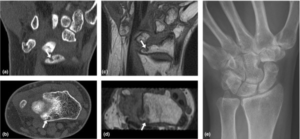
Background: The objectives of the present study were, with multidetector computed tomography (CT) as the reference method, to determine the performance of magnetic resonance imaging (MRI) and radiography for the detection of bone erosions in rheumatoid arthritis wrist bones, and to test whether measuring volumes of erosions on CT and MRI is reproducible and correlated to semiquantitative assessments (scores) of erosions on CT, MRI and radiography.
Methods: Seventeen patients with rheumatoid arthritis and four healthy control individuals underwent CT, MRI and radiography of one wrist, performed on the same day. CT was performed on a Philips Mx8000IDT unit (voxel size 0.4 mm x 0.4 mm x 1 mm) and MRI was performed on a Philips Panorama 0.6T unit (voxel size 0.4 mm x 0.4 mm x 0.4 mm). Images were evaluated separately for erosions in all wrist bones and were scored according to the principles of the Outcome Measures in Rheumatology Rheumatoid Arthritis MRI Scoring System (CT and MRI) and the Sharp/van der Heijde (radiographs) scoring methods. Measurements of erosion volumes of all erosions were performed twice with a 1-week interval.
Results: With CT as the reference method, the overall sensitivity, specificity and accuracy (concordance) of MRI for detecting erosions were 61%, 93% and 77%, respectively, while the respective values were 24%, 99% and 63% for radiography. The intramodality agreements when measuring erosion volumes were high for both CT and MRI (Spearman correlation coefficients 0.92 and 0.90 (both P < 0.01), respectively). Correlations between volumes and scores of individual erosions were 0.96 for CT and 0.99 for MRI, while they were 0.83 (CT) and 0.80 (MRI) for persons' total erosion volume and total score (all P < 0.01).
Conclusion: With CT as the reference method, MRI showed moderate sensitivity and good specificity and accuracy for detection of erosions in rheumatoid arthritis and healthy wrist bones, while radiography showed very low sensitivity. The tested volumetric method was highly reproducible and correlated to scores of erosions.
Figures
Similar articles
-
Validity of a computer-assisted manual segmentation software to quantify wrist erosion volume using computed tomography scans in rheumatoid arthritis.BMC Musculoskelet Disord. 2013 Sep 12;14:265. doi: 10.1186/1471-2474-14-265. BMC Musculoskelet Disord. 2013. PMID: 24028158 Free PMC article.
-
The smallest detectable difference and sensitivity to change of magnetic resonance imaging and radiographic scoring of structural joint damage in rheumatoid arthritis finger, wrist, and toe joints: a comparison of the OMERACT rheumatoid arthritis magnetic resonance imaging score applied to different joint combinations and the Sharp/van der Heijde radiographic score.Arthritis Rheum. 2005 Aug;52(8):2300-6. doi: 10.1002/art.21207. Arthritis Rheum. 2005. PMID: 16052593
-
Rheumatoid arthritis bone erosion volumes on CT and MRI: reliability and correlations with erosion scores on CT, MRI and radiography.Ann Rheum Dis. 2007 Oct;66(10):1388-92. doi: 10.1136/ard.2007.072520. Epub 2007 Jul 2. Ann Rheum Dis. 2007. PMID: 17606464 Free PMC article.
-
Imaging of the hand and wrist in RA.Ann Rheum Dis. 2002 Oct;61(10):867-9. doi: 10.1136/ard.61.10.867. Ann Rheum Dis. 2002. PMID: 12228153 Free PMC article. Review. No abstract available.
-
[Imaging modalities in rheumatoid arthritis].Ryumachi. 1997 Aug;37(4):587-99. Ryumachi. 1997. PMID: 9311286 Review. Japanese. No abstract available.
Cited by
-
[Rheumatoid arthritis of the hand : Part 2: Imaging].Radiologe. 2021 Apr;61(4):362-374. doi: 10.1007/s00117-021-00833-3. Epub 2021 Mar 16. Radiologe. 2021. PMID: 33728480 Review. German.
-
Imaging in rheumatoid arthritis: the role of magnetic resonance imaging and computed tomography.Radiol Med. 2019 Nov;124(11):1128-1141. doi: 10.1007/s11547-019-01014-y. Epub 2019 Mar 18. Radiol Med. 2019. PMID: 30880357 Review.
-
Carpal pseudoerosions: a plain X-ray interpretation pitfall.Skeletal Radiol. 2014 Oct;43(10):1377-85. doi: 10.1007/s00256-014-1907-5. Epub 2014 Jun 6. Skeletal Radiol. 2014. PMID: 24902509
-
Joint blood flow is more sensitive to inflammatory arthritis than oxyhemoglobin, deoxyhemoglobin, and oxygen saturation.Biomed Opt Express. 2016 Sep 1;7(10):3843-3854. doi: 10.1364/BOE.7.003843. eCollection 2016 Oct 1. Biomed Opt Express. 2016. PMID: 27867697 Free PMC article.
-
Diagnostic performance of magnetic resonance imaging for detecting osseous abnormalities of the temporomandibular joint and its correlation with cone beam computed tomography.Dentomaxillofac Radiol. 2010 Jul;39(5):270-6. doi: 10.1259/dmfr/25151578. Dentomaxillofac Radiol. 2010. PMID: 20587650 Free PMC article.
References
-
- Boers M, Felson DT. Clinical measures in rheumatoid arthritis: which are most useful in assessing patients? J Rheumatol. 1994;21:1773–1774. - PubMed
-
- van der Heijde DM, van Leeuwen MA, van Riel PL, Koster AM, van't Hof MA, van Rijswijk MH, van de Putte LB. Biannual radiographic assessments of hands and feet in a three-year prospective followup of patients with early rheumatoid arthritis. Arthritis Rheum. 1992;35:26–34. doi: 10.1002/art.1780350105. - DOI - PubMed
Publication types
MeSH terms
LinkOut - more resources
Full Text Sources
Medical
