. 2008 Mar;82(3):673-84.
doi: 10.1016/j.ajhg.2007.12.019. Epub 2008 Feb 28.

Array-based gene discovery with three unrelated subjects shows SCARB2/LIMP-2 deficiency causes myoclonus epilepsy and glomerulosclerosis

Affiliations

Array-based gene discovery with three unrelated subjects shows SCARB2/LIMP-2 deficiency causes myoclonus epilepsy and glomerulosclerosis

Samuel F Berkovic et al. Am J Hum Genet. 2008 Mar.

Abstract

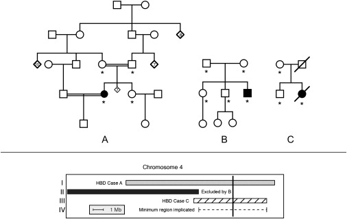
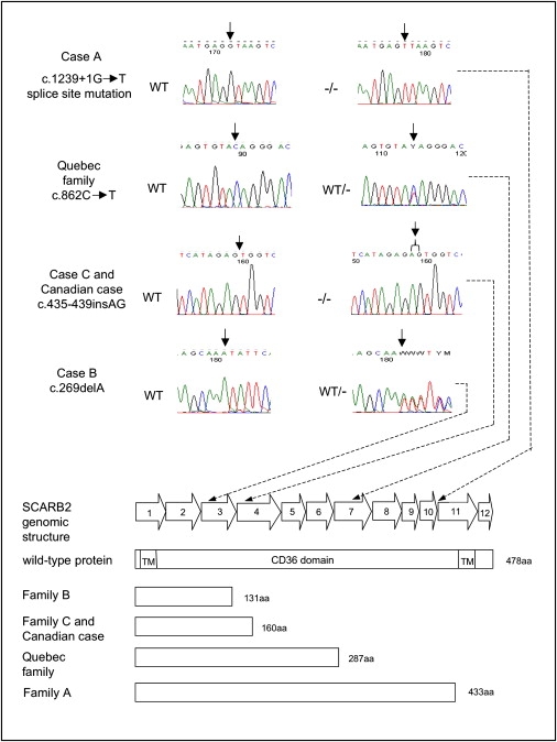
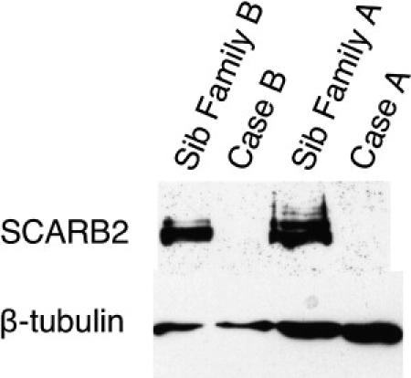
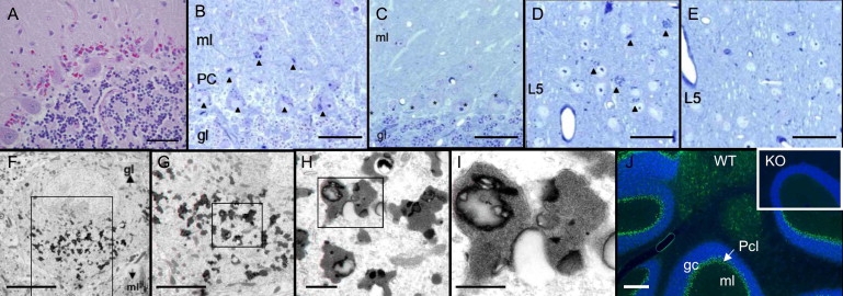
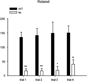
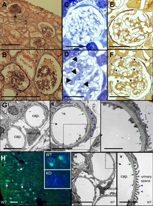
Action myoclonus-renal failure syndrome (AMRF) is an autosomal-recessive disorder with the remarkable combination of focal glomerulosclerosis, frequently with glomerular collapse, and progressive myoclonus epilepsy associated with storage material in the brain. Here, we employed a novel combination of molecular strategies to find the responsible gene and show its effects in an animal model. Utilizing only three unrelated affected individuals and their relatives, we used homozygosity mapping with single-nucleotide polymorphism chips to localize AMRF. We then used microarray-expression analysis to prioritize candidates prior to sequencing. The disorder was mapped to 4q13-21, and microarray-expression analysis identified SCARB2/Limp2, which encodes a lysosomal-membrane protein, as the likely candidate. Mutations in SCARB2/Limp2 were found in all three families used for mapping and subsequently confirmed in two other unrelated AMRF families. The mutations were associated with lack of SCARB2 protein. Reanalysis of an existing Limp2 knockout mouse showed intracellular inclusions in cerebral and cerebellar cortex, and the kidneys showed subtle glomerular changes. This study highlights that recessive genes can be identified with a very small number of subjects. The ancestral lysosomal-membrane protein SCARB2/LIMP-2 is responsible for AMRF. The heterogeneous pathology in the kidney and brain suggests that SCARB2/Limp2 has pleiotropic effects that may be relevant to understanding the pathogenesis of other forms of glomerulosclerosis or collapse and myoclonic epilepsies.


Figures

Figure 1
Figure 1
Linkage Analysis Shown in the upper panel: pedigrees used to map AMRF. Families A and B were previously clinically described as families 2 and 1, respectively (3), and pedigree C here was family B in . Filled symbols indicate cases with AMRF. Asterisks indicate individuals used for genotyping. Shown in the lower panel: localization of the critical region on chromosome 4 (see text). Region I (gray bar) was identified as homozygous by descent (HBD) in case A and not shared by the sibling of case A. In region II, case B shared both haplotypes identical by descent with unaffected siblings, thus excluding this region. Region III shows the homozygous region of case C. Region IV (dashed line) shows the deduced critical region of 6.6 Mb. The vertical black line indicates the position of SCARB2.
Figure 2
Figure 2
SCARB2 Mutations Genomic structure of the SCARB2 gene and the position of the mutations found in the AMRF cases. The sequencing chromatograms indicate the mutations found in the affected individuals or in a carrier, and the predicted effects of the mutations on the SCARB2 protein are illustrated below. “WT” refers to an individual lacking a mutation, “WT/−” refers to a heterozygote, and “−/−” refers to an individual homozygous for a mutation.
Figure 3
Figure 3
Western Blots with Lymphoblastoid Cell Lines from Cases A and B and Their Unaffected Siblings SCARB2 was probed with a polyclonal goat antibody to Limp2 and is shown in the samples from the two siblings as a band at 75–80 kD; it was absent in the two cases. β-tubulin is shown in all four subjects at ∼50kD as a control.
Figure 4
Figure 4
Neuropathology of AMRF and Limp2-Deficient Mice (A) Human cerebellar vermis (autopsy sample; case C). Magenta-colored granules are seen between intact Purkinje cells (PAS stain; the scale bar represents 0.05 mm). (B–E) Light microscopy of Limp2−/− brain in 16-month-old mice (semithin sections, toluidine blue). Cerebellar cortex of Limp2−/− mouse (B) shows all Purkinje cells (PC) as well as some neurons and glial cells in the molecular layer (ml) with numerous cytoplasmic inclusions (dense bodies) (arrowheads). In control wild-type mice (C), no inclusions are seen in the Purkinje cells (marked with asterisks), granular layer (gl), or ml. Sections of cerebral cortex from Limp2−/− show similar cytoplasmic inclusions that are in neuronal perikarya of laminae 2/3 and 5 (D) and that are absent in the cerebral cortex of wild-type mice (E). Layer 5 (L5) is shown. Scale bars represent 50 μm. (F–I) Electron microscopy of Limp2−/− mouse brain. One Purkinje cell shown at increasing magnifications from left to right. The boxes indicate the region shown at higher magnification. The osmiophilic inclusions were most frequently observed within the perikarya and only very rarely within dendrites (not shown). The membrane-limited inclusions contain homogeneously granular material, some lipid droplets, and occasionally some lamellar structures. Scale bars represent 10 μm, 5 μm, 1 μm, and 0.5 μm, respectively. (J) Immunohistological studies of mouse cerebellum. Wild-type cerebellar cortex (WT) is shown with nuclei stained with DAPI in blue. Limp2 is shown in green and is concentrated in the Purkinje cell layer (the scale bar represents 200 μm). The inset shows knockout (KO) with absence of Limp2 in the Purkinje cell layer.
Figure 5
Figure 5
Rotarod Testing of Limp2−/− and Wild-Type Mice Accelerating rotarod performance in wild-type (black bars) and Limp2−/− (white bars) mice. On each of the 5 min trials, Limp2−/− mice were unable to balance on the rod quite as long as wild-type (control) mice, and this demonstrates the ataxia and neuromotor impairment in the Limp2-deficient group. Data are means and SEM; asterisks indicate significance of difference with control values (post hoc Fisher LSD), p < 0.05, ∗∗p < 0.01, and ∗∗∗p = 0.001.
Figure 6
Figure 6
Glomerular Pathology in AMRF and Limp2-Deficient Mice (A and B) Human glomerular pathology. Representative renal cortex (A) of human case C with AMRF showing an almost totally sclerosed glomerulus (arrow) because of global collapse and two glomeruli showing the early phases of collapse and sclerosis. Higher power (B) of the two glomeruli in the early phases of capillary collapse with partially obliterated lumina and prominent epithelial cells caused by hypertrophy and hyperplasia (silver methernamine-Marson's trichrome) is shown. Scale bars represent 50 μm. (C–F) Light microscopy of Limp2−/− mice. Glomerulus of a wild-type (WT) mouse aged 16 months with the typical tissue structure (C) compared to Limp2−/− (KO) mouse (D), with arrowheads indicating an expanded messangium (semithin sections, toluidine blue). The same wild-type animal with silver stain for highlighting extracellular matrix and basal membranes (E) compared to the Limp2−/− mouse (F) with a mesangium that is expanded (arrowheads) is shown. Scale bars represent 50 μm. (G) Electron microscopy of Limp2−/− (Gi–Giii) and wild-type (G iv–Gv) mice. Images show effacement of the foot processes of the podocytes (blue arrowheads) and thickening of the glomerular basal membrane (green arrows) in Limp2−/− (KO) mice compared to normal podocyte morphology and basement-membrane thickness in wild-type (WT) animals. Scale bars represent 10 μm (Gi and Giv), 5 μm (Gii), and 2.5 μm (Giii and Gv). (H) Immunohistological studies of mouse renal cortex. The overview shows wild-type cortex with nuclei in blue (DAPI) and Limp2 in green; glomeruli are indicated by arrows (scale bar represents 200 μm). High-magnification insets of glomeruli show Limp2 in wild-type animals but absent in knockout animals.

References

    1. Andermann E., Andermann F., Carpenter S., Wolfe L.S., Nelson R., Patry G., Boileau J. Action myoclonus-renal failure syndrome: A previously unrecognized neurological disorder unmasked by advances in nephrology. Adv. Neurol. 1986;43:87–103. - PubMed
    1. Badhwar A., Berkovic S.F., Dowling J.P., Gonzales M., Narayanan S., Brodtmann A., Berzen L., Caviness J., Trenkwalder C., Winkelmann J. Action myoclonus-renal failure syndrome: Characterization of a unique cerebro-renal disorder. Brain. 2004;127:2173–2182. - PubMed
    1. Vadlamudi L., Vears D.F., Hughes A., Pedagogus E., Berkovic S.F. Action myoclonus-renal failure syndrome: A cause for worsening tremor in young adults. Neurology. 2006;67:1310–1311. - PubMed
    1. Di X., Matsuzaki H., Webster T.A., Hubbell E., Liu G., Dong S., Bartell D., Huang J., Chiles R., Yang G. Dynamic model based algorithms for screening and genotyping over 100 K SNPs on oligonucleotide microarrays. Bioinformatics. 2005;21:1958–1963. - PubMed
    1. Terwilliger J.D., Ott J. Johns Hopkins University Press; Baltimore, London: 1994. Handbook of Human Genetic Linkage.

Publication types

Substances