

The right parietal lobe is critical for visual working memory
- PMID: 18308348
- PMCID: PMC2441642
- DOI: 10.1016/j.neuropsychologia.2008.01.009
The right parietal lobe is critical for visual working memory
Abstract
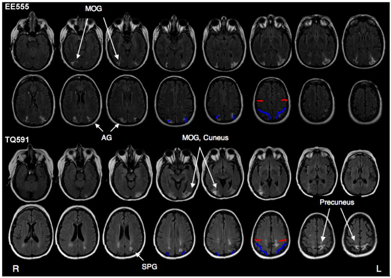
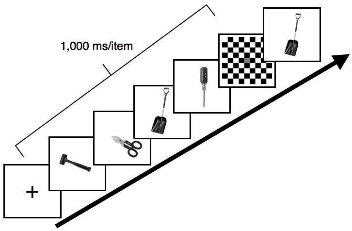
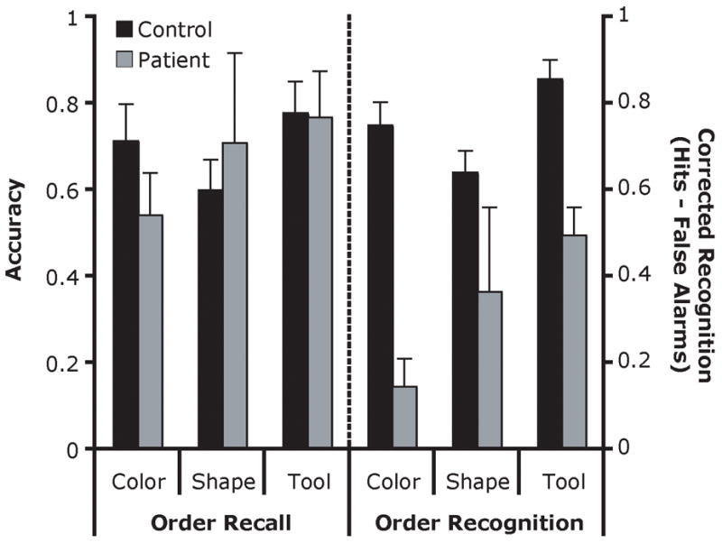
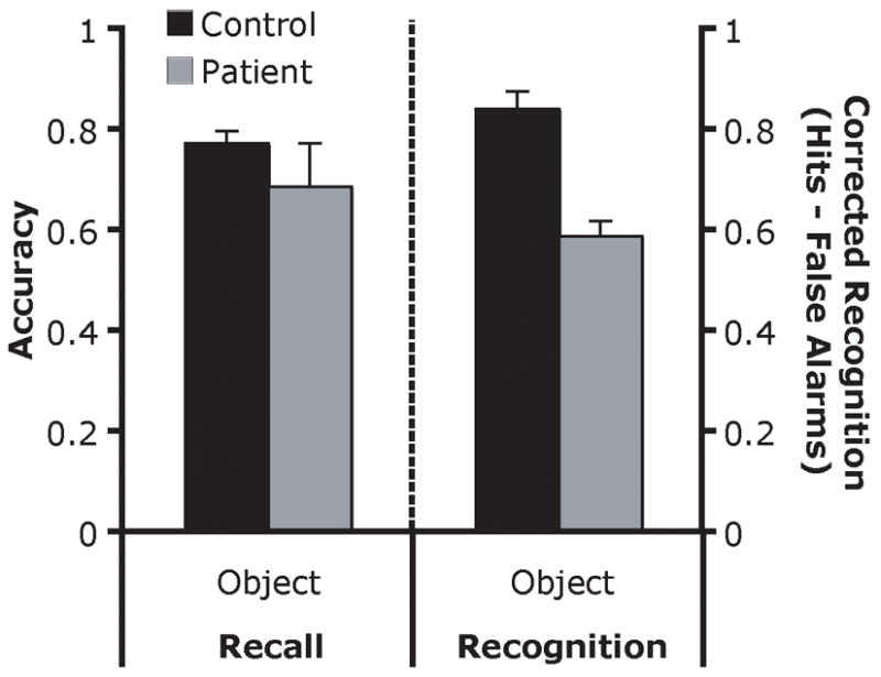
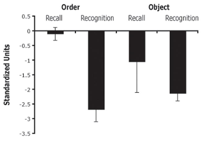
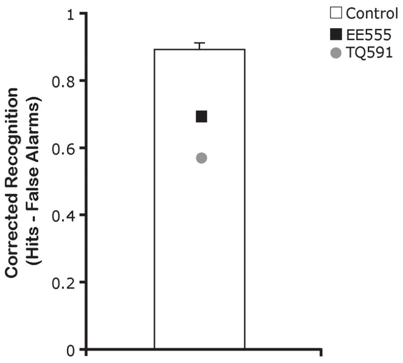
Visual working memory (VWM) permits the maintenance of object identities and their locations across brief delays such as those accompanying eye movements. Recent neuroimaging studies have emphasized the role of the posterior parietal lobe in this process although the specific nature of this involvement in VWM remains controversial. Neuroimaging findings suggest that the parietal lobe may have a general role in remembering various types of visual information whereas neuropsychological findings suggest that parietal involvement is primarily related to motor spatial attention and spatial memory. In the present study, patients with unilateral right parietal lobe damage, lacking symptoms of neglect, were tested in several VWM old/new recognition tasks. Parietal damage lead to impaired performance on all VWM tasks, including spatial, object, and object/spatial conjunction tasks. Deficits were found across several stimulus categories. These results provide neuropsychological support for neuroimaging results, and more generally indicate that the parietal lobe serves a general role in diverse forms of VWM.
Figures
References
-
- Aggleton JP, Brown MW. Episodic memory, amnesia, and the hippocampal-anterior thalamic axis. Behav Brain Sci. 1999;22(3):425–444. discussion 444–489. - PubMed
-
- Aggleton JP, Shaw C. Amnesia and recognition memory: a re-analysis of psychometric data. Neuropsychologia. 1996;34(1):51–62. - PubMed
-
- Awh E, Barton B, Vogel EK. Visual working memory represents a fixed number of items regardless of complexity. Psychol Sci. 2007;18:622–628. - PubMed
-
- Baldo JV, Dronkers NF. The role of inferior parietal and inferior frontal cortex in working memory. Neuropsychology. 2006;20(5):529–538. - PubMed
-
- Basso A, Spinnler H, Vallar G, Zanobio ME. Left hemisphere damage and selective impairment of auditory verbal short-term memory. A case study. Neuropsychologia. 1982;20(3):263–274. - PubMed
Publication types
MeSH terms
Grants and funding
LinkOut - more resources
Full Text Sources
Medical
