. 2008 May 25;375(1):1-12.
doi: 10.1016/j.virol.2008.01.042. Epub 2008 Mar 4.

APOBEC3G restricts early HIV-1 replication in the cytoplasm of target cells

Affiliations

APOBEC3G restricts early HIV-1 replication in the cytoplasm of target cells

Jenny L Anderson et al. Virology. .

Abstract

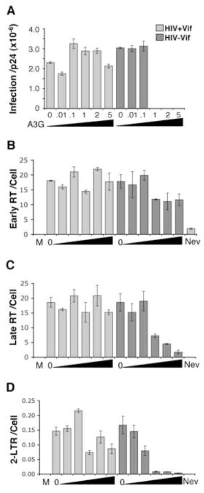
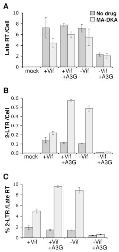
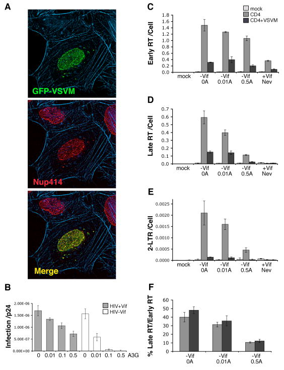
Cellular APOBEC3G (A3G) protein is packaged into human immunodeficiency virus type 1 (HIV-1) virions in producer cells yet restricts viral replication in target cells. To characterize this restriction in target cells, the effect of A3G on generating various HIV-1 cDNA products was measured by quantitative real-time PCR. A3G decreased cDNA products from Vif-deficient HIV-1, with minor effects on early reverse transcripts and larger declines in late reverse transcripts. However, the greatest decline was typically observed in nuclear 2-LTR circles. Moreover, the magnitude of these declines varied with A3G dose. Adding integration inhibitor did not stop the A3G-mediated loss in 2-LTR circles. Moreover, obstructing HIV-1 nuclear entry using vesicular stomatitis virus matrix protein did not stop the A3G-mediated decline in late reverse transcripts. Collectively, these data suggest that A3G has important restriction activity in the cytoplasm and progressively diminishes viral cytoplasmic and nuclear cDNA forms with increasing magnitude during restriction.


Figures

Figure 1
Figure 1. Human A3G impairs HIV-1ΔVif 2-LTR circle formation
(A) Human A3G impairs the infectivity of VSVg pseudotyped HIV-1 lacking Vif but not Vif positive HIV-1. VSVg pseudotyped HIV-1 or HIV-1ΔVif were produced with different human A3G amounts, as indicated on the graph by the molar ratio of A3G to provirus plasmid transfected. The infectivity of this virus on MAGI reporter cells, measured using a liquid β-gal assay (infection), per p24Gag in the virus stock (p24) is shown for a representative experiment. Error bars indicate standard deviation in triplicate values. HIV-1 without VSVg envelope acts a negative control. (B–D) A3G potently impairs 2-LTR circle production from HIV-1ΔVif. HOS cells were continuously infected 15 hours (h) with 7.94 ng of VSVg pseudotyped HIV-1 without or with A3G (2:1 molar ratio). The virus was removed, new media added and the intracellular DNA harvested at 15 h, 26 h or 48 h from the start of infection (light, medium and dark grey bars respectively). Viral early RT (B), late RT (C), 2-LTR circle (D) and cellular β-actin were quantified by real-time PCR and values shown per cell (β-actin). Error bars indicate standard deviation in triplicate values from a representative experiment. The negligible viral cDNA in cells infected with virus plus the Nevirapine (Nev) reverse transcription inhibitor, demonstrates viral reverse transcription products are measured in this assay.
Figure 2
Figure 2. Primers with or without A3G hypermutation sites show similar decreases in HIV-1ΔVif cDNA products by A3G
(A) Primer sequences prone (+) or not prone (−) to A3G hypermutation and used to amplify 2-LTR circle (upper) and late RT (lower) viral cDNA products are shown. F, forward (plus strand) primer; R, reverse (minus strand) primer. Dark and light grey boxes indicate sequence sensitive to A3G hypermutation at high and low frequency respectively (Yu et al., 2004b). (B–C) HOS cells were continuously infected for 14 h with 3.42 ng of HIV-1 or HIV-1ΔVif lacking or containing A3G (made at 1:1 molar ratio of A3G:provirus plasmid). Intracellular DNA was analysed for 2-LTR circles (B) or late RT (C) using primers containing sequence prone (dark grey) or not prone (light grey) to high frequency A3G hypermutation.
Figure 3
Figure 3. Declines in HIV-1 cDNA products vary with A3G dose
(A) VSVg pseudotyped HIV-1 with (light grey) or without (dark grey) Vif was produced with increasing levels of human A3G (indicated by the molar ratio of A3G per 1 provirus plasmid plus increasing size of the black triangle). The infectivity of these virus stocks on MAGI reporter cells per virion p24Gag concentration is shown for a representative experiment. (B–D) HOS cells were continuously infected with 29.77 ng of these viruses for 18 h and intracellular DNA measured for viral early RT (B), late RT (C) or 2-LTR circles (D) per cell (β-actin) using real-time PCR. Error bars indicate standard deviation of triplicate values. M, mock (no virus); 0, virus without A3G; Nev, Nevirapine. Increasing A3G during HIV-1ΔVif production correlates with larger declines in late RT and 2-LTR circles in target cells.
Figure 4
Figure 4. MA-DKA integration inhibitor reveals human A3G impairs HIV-1 provirus as well as 2-LTR circles
HOS cells were pretreated 4 h with MA-DKA (light grey) or DMSO (medium grey) as a no drug control and continuously infected 24 h with 16.27 ng of VSVg pseudotyped HIV-1 produced without or with A3G (2:1 molar ratio of A3G:provirus plasmid). Intracellular DNA was analysed for late RT (A) or 2-LTR circles (B) per cell (β-actin) by real-time PCR, with the percentage of 2-LTR circles per late RT also shown to account for changes in late RT between samples (C). Error bars indicate standard deviation in triplicate values from a representative experiment. As 2-LTR circle output from HIV-1ΔVif plus A3G remains poor with MA-DKA integration inhibitor indicates A3G impedes HIV-1ΔVif provirus as well as 2-LTR circle forms.
Figure 5
Figure 5. A3G still depletes HIV-1 Late RT when VSV matrix protein impairs nuclear entry
(A) HeLa cells 24 h after transfection with pEGFP-VSVM (green), and stained for nuclear pore complexes using anti-Nup414 nucleoporin antibody (red) and filamentous actin using texas-red phalloidin (blue). GFP-VSVM localizes to nuclear pore complexes. (B) HIV-1 with (grey) or without (white) Vif and containing wildtype envelope protein were produced with increasing levels of human A3G (molar ratio of A3G to 1 provirus plasmid shown). The infectivity of these virus stocks on MAGI reporter cells per p24Gag concentration is shown for a representative experiment. (C–F) A, A3G; Mock, no virus; Nev, Nevirapine. HeLa cells were transfected without (light grey), with pCD4 (medium grey) or with pEGFP-VSVM plus pCD4 (dark grey) and 24 h later, continuously infected with 36.74 ng of the above HIV-1ΔVif stocks a further 24 h. Intracellular DNA was then analysed for viral early RT (C), late RT (D) or 2-LTR circles (E) per cell, the latter acting as a surrogate for nuclear localization of viral cDNA. The % of late RT per early RT is also shown (F) to demonstrate when changes in early RT levels are accounted for, A3G still decreases HIV-1 late RT when viral cDNA import is impaired by VSVM. Error bars indicate standard deviation of triplicate values.

References

    1. Aires da Silva F, Santa-Marta M, Freitas-Vieira A, Mascarenhas P, Barahona I, Moniz-Pereira J, Gabuzda D, Goncalves J. Camelized rabbit-derived VH single-domain intrabodies against Vif strongly neutralize HIV-1 infectivity. J Mol Biol. 2004;340(3):525–42. - PubMed
    1. Bishop KN, Holmes RK, Malim MH. Antiviral potency of APOBEC proteins does not correlate with cytidine deamination. J Virol. 2006;80(17):8450–8. - PMC - PubMed
    1. Bishop KN, Holmes RK, Sheehy AM, Davidson NO, Cho SJ, Malim MH. Cytidine deamination of retroviral DNA by diverse APOBEC proteins. Curr Biol. 2004;14(15):1392–6. - PubMed
    1. Bogerd HP, Wiegand HL, Doehle BP, Lueders KK, Cullen BR. APOBEC3A and APOBEC3B are potent inhibitors of LTR-retrotransposon function in human cells. Nucleic Acids Res. 2006a;34(1):89–95. - PMC - PubMed
    1. Bogerd HP, Wiegand HL, Hulme AE, Garcia-Perez JL, O’Shea KS, Moran JV, Cullen BR. Cellular inhibitors of long interspersed element 1 and Alu retrotransposition. Proc Natl Acad Sci U S A. 2006b;103(23):8780–5. - PMC - PubMed

Publication types