. 2008 Mar 11;105(10):3903-8.
doi: 10.1073/pnas.0712321105. Epub 2008 Feb 28.

Suppression of non-small cell lung tumor development by the let-7 microRNA family

Affiliations

Suppression of non-small cell lung tumor development by the let-7 microRNA family

Madhu S Kumar et al. Proc Natl Acad Sci U S A. .

Abstract

Many microRNAs (miRNAs) target mRNAs involved in processes aberrant in tumorigenesis, such as proliferation, survival, and differentiation. In particular, the let-7 miRNA family has been proposed to function in tumor suppression, because reduced expression of let-7 family members is common in non-small cell lung cancer (NSCLC). Here, we show that let-7 functionally inhibits non-small cell tumor development. Ectopic expression of let-7g in K-Ras(G12D)-expressing murine lung cancer cells induced both cell cycle arrest and cell death. In tumor xenografts, we observed significant growth reduction of both murine and human non-small cell lung tumors when overexpression of let-7g was induced from lentiviral vectors. In let-7g expressing tumors, reductions in Ras family and HMGA2 protein levels were detected. Importantly, let-7g-mediated tumor suppression was more potent in lung cancer cell lines harboring oncogenic K-Ras mutations than in lines with other mutations. Ectopic expression of K-Ras(G12D) largely rescued let-7g mediated tumor suppression, whereas ectopic expression of HMGA2 was less effective. Finally, in an autochthonous model of NSCLC in the mouse, let-7g expression substantially reduced lung tumor burden.


Conflict of interest statement

The authors declare no conflict of interest.

Figures
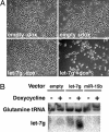
Fig. 1.
Fig. 1.
Let-7g impairs proliferation and enhances cell death. (A) LKR13-Tet-On-KRAB-TE-empty and -let-7g cells were plated (5 × 10⁵ cells per plate). Twelve hours later, cells were placed in the presence/absence of 5 μg/ml doxycycline. Forty-eight hours later, images were taken by phase contrast microscopy. (B) Small RNA Northern blot analysis was performed against let-7g and Glutamine tRNA in LKR13-Tet-On-KRAB-TE-empty, -miR-15b, and -let-7g (see SI Text for details) cells in the presence or absence of 5 μg/ml doxycycline.
Fig. 2.
Fig. 2.
Let-7g suppresses tumorigenesis in vivo. (A) LKR13-Tet-On-KRAB-TE-empty, -miR-15b, and -let-7g cells were plated in the presence of 5 μg/ml doxycycline. Twenty-four hours later, cells were sorted and injected s.c. into immune-compromised mice (2.5 × 10⁴ cells per injection). Two days later, mice were treated with drinking water containing doxycycline (2 mg/ml) and sucrose (4% wt/vol), and tumor values were measured over time. Values are mean ± SEM (n = 6). (B) LKR13-Tet-On-KRAB-TE-let-7g cells were treated with doxycycline, sorted, and injected as described above. Two days later, mice were treated with either drinking water containing doxycycline (2 mg/ml) and sucrose (4% wt/vol) or drinking water containing sucrose alone. Tumor values were measured over time. Values are mean ± SEM (n = 6). (C–E) Tet-On-KRAB-TE-let-7g cells were generated in A549 (C), Calu-1 (D), and H1650 (E) cells. Cells were prepared and injected (106 cells per injection) into immune-compromised mice. Mice were treated and tumors were measured as above. Values are mean ± SEM (n = 6).
Fig. 3.
Fig. 3.
Let-7g-induced tumors maintain overexpression and target suppression. (A) Small RNA Northern blotting was performed against let-7g and Glutamine tRNA in LKR13-Tet-On-KRAB-TE-empty and -let-7g tumors generated from mice treated with or without doxycycline in the drinking water as described above. (B) Western blot analysis was performed in LKR13-Tet-On-KRAB-TE-empty and -let-7g tumors generated from mice treated with doxycycline in the drinking water as described above.
Fig. 4.
Fig. 4.
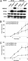
K-RasG12D substantially rescues let-7g-mediated tumor suppression. (A) LKR13-Tet-On-KRAB-TE-miR-15b and -let-7g cells were infected with pBabe.Zeo.empty, K-RasG12D, and HMGA2 and Western blot analysis was performed. (B and C) LKR13-Tet-On-KRAB-TE-let-7g cells infected with pBabe.Zeo.K-RasG12D (B) or HMGA2 (C) were prepared and injected (106 cells per injection) into immune-compromised mice. Mice were treated and tumors were measured as above. Values are mean ± SEM (n = 6).
Fig. 5.
Fig. 5.
Let-7g suppresses tumor initiation in an autochthonous NSCLC model. (A) Diagram of the Puro.Cre lentiviral vector for coexpression of let-7g/let-7g sm with Cre recombinase. (B) Small RNA Northern blot analysis was performed against let-7g (both wild type and seed mutant), miR-17 and Glutamine tRNA in HEK293 cells infected with Puro.Cre (empty), Puro.let7gsm.Cre (let-7g sm), and Puro.let7g.Cre (let-7g). (C–E) KrasLSL-G12D;Trp-53flox/flox mice were intratracheally infected with the Puro.Cre lentiviral vectors described above. Twelve weeks after infection, animals were killed, and tumor number (C), tumor and lung area (D), and tumor size (E) were quantified with Bioquant software. Values are mean ± SEM (n = 9 for empty, n = 11 for let-7g, and n = 11 for let-7g sm). *, P < 0.0005, #, P < 0.01; ###, P < 0.1. (F) Small RNA Northern blotting was performed against let-7g (both wild type and seed mutant), and Glutamine tRNA on lung tumors was generated from KrasLSL-G12D;Trp-53flox/flox mice infected with Puro.Cre (empty), Puro.let7g.Cre (let-7g), and Puro.let7gsm.Cre (let-7g sm). (G) Western blot analysis was performed on lung tumors generated from KrasLSL-G12D;Trp-53flox/flox mice infected with Puro.Cre (empty), Puro.let7g.Cre (let-7g), and Puro.let7gsm.Cre (let-7g sm). All samples were probed on the same blot.

Similar articles

Cited by

References

    1. Bartel DP. MicroRNAs: genomics, biogenesis, mechanism, and function. Cell. 2004;116:281–297. - PubMed
    1. Valencia-Sanchez MA, Liu J, Hannon GJ, Parker R. Control of translation and mRNA degradation by miRNAs and siRNAs. Genes Dev. 2006;20:515–524. - PubMed
    1. Lewis BP, Burge CB, Bartel DP. Conserved seed pairing, often flanked by adenosines, indicates that thousands of human genes are microRNA targets. Cell. 2005;120:15–20. - PubMed
    1. Lewis BP, Shih IH, Jones-Rhoades MW, Bartel DP, Burge CB. Prediction of mammalian microRNA targets. Cell. 2003;115:787–798. - PubMed
    1. Calin GA, et al. Human microRNA genes are frequently located at fragile sites and genomic regions involved in cancers. Proc Natl Acad Sci USA. 2004;101:2999–3004. - PMC - PubMed

Publication types