. 2008 Mar 11;105(10):4044-9.
doi: 10.1073/pnas.0710294105. Epub 2008 Feb 28.

A SPIKE1 signaling complex controls actin-dependent cell morphogenesis through the heteromeric WAVE and ARP2/3 complexes

Affiliations

A SPIKE1 signaling complex controls actin-dependent cell morphogenesis through the heteromeric WAVE and ARP2/3 complexes

Dipanwita Basu et al. Proc Natl Acad Sci U S A. .

Abstract

During morphogenesis, the actin cytoskeleton mediates cell-shape change in response to growth signals. In plants, actin filaments organize the cytoplasm in regions of polarized growth, and the filamentous arrays can be highly dynamic. Small GTPase signaling proteins termed Rho of plants (ROP)/RAC control actin polymerization. ROPs cycle between inactive GDP-bound and active GTP-bound forms, and it is the active form that interacts with effector proteins to mediate cytoskeletal rearrangement and cell-shape change. A class of proteins termed guanine nucleotide exchange factors (GEFs) generate GTP-ROP and positively regulate ROP signaling. However, in almost all experimental systems, it has proven difficult to unravel the complex signaling pathways from GEFs to the proteins that nucleate actin filaments. In this article, we show that the DOCK family protein SPIKE1 (SPK1) is a GEF, and that one function of SPK1 is to control actin polymerization via two heteromeric complexes termed WAVE and actin-related protein (ARP) 2/3. The genetic pathway was constructed by using a combination of highly informative spk1 alleles and detailed analyses of spk1, wave, and arp2/3 single and double mutants. Remarkably, we find that in addition to providing GEF activity, SPK1 associates with WAVE complex proteins and may spatially organize signaling. Our results describe a unique regulatory scheme for ARP2/3 regulation in cells, one that can be tested for widespread use in other multicellular organisms.


Conflict of interest statement

The authors declare no conflict of interest.

Figures

Fig. 1.
Fig. 1.
The SPK1 Dock homology region 2 (DHR2) domain provides critical functions in vivo. (A) The domain organization of SPK1 protein. The locations of the spk1-1 and spk1-3 mutations are indicated. The conserved regions of SPK1 are labeled. (B) Wild-type, spk1-1, and spk1-3 seedlings at 10 DAG. (C) Pavement cell–cell adhesion defects of spk1 cotyledons and leaves. Wild-type, spk1-1, and spk1-3 seedlings were stained with 0.1 μM FM1–43 (green) to visualize cell boundaries. The autofluorescence from the chloroplasts of the underlying mesophyll cells is red and is clearly visible within pavement cell gaps. (D) SEM of stage 6 mature trichomes from wild-type, spk1-1, and spk1-3 leaves, respectively. (E) The truncated spk1-3 accumulates. (Upper) Lanes 1–4, Western blot analysis of protein of whole-cell extracts of wild-type, spk1-1, spk1-2, and spk1-3 plants, respectively, probed with α-SPK1N antibody. (Lower) Western blot analysis with same extracts as in Upper but probed with a α-PEPC antibody for a loading control. [Scale bars: 1 cm (B), 10 μm (C), and 100 μm (D).]
Fig. 2.
Fig. 2.
SPK1 binds to ROPs in a DHR2-dependent manner and facilitates nucleotide exchange. (A) Endogenous SPK1–ROP complexes require an intact DHR2 domain. Lanes 1 and 5, 5% of input solubilized protein extracts; lanes 2–4, proteins immunoprecipitated from wild-type extracts using α-SPK1N antibody, preimmune serum, and protein A beads alone, respectively; lanes 6–8, same conditions as above but with spk1-3 extracts. coIP fractions were probed with α-SPK1N (Upper) and α-ROP (Lower) antibodies. (B) Endogenous SPK1 associates specifically with inactive forms of ROP2 in a DHR2-dependent manner. GTP-bound, GDP-bound, or nucleotide-depleted (Nt-D) GST–ROP2 and human GST–Rac1 were bound to beads and incubated with cytosolic fractions from wild-type (Upper) and spk1-3 (Lower) plants. Bound fractions were probed with α-SPK1N antibodies. (C) Recombinant purified 996-DHR2 interacts directly with ROP in a nucleotide-dependent manner. Lane 1, 10% of the input fraction; lanes 2–4, the GST-996-DHR2 bound fractions of GTP-loaded, GDP-loaded, or Nt-D HIS–ROP2, respectively. Bound fractions were probed with an α-ROP antibody. (D) Recombinant full-length SPK1 interacts directly with inactive ROPs. GTP-bound, GDP-bound, or Nt-D GST–ROPs were used in GST pull-down assays with recombinant full-length SPK1 expressed in insect SF9 cells. Lane 1, 10% of total input SPK1; lanes 2–25, Western blots of bound, pellet fractions probed with the α-SPK1N antibody. (E) Full-length SPK1 has guanine nucleotide exchange activity in vitro. Purified ROP2 was loaded with [3H]GDP, and exchange reactions were initiated by the addition of 100-fold molar excess of unlabeled GTP and SPK1. Controls included no additional proteins and SF9 extracts containing an irrelevant protein. Each time point is shown as the mean ± SD of triplicate measurements. (F) The 996-DHR2 fragment of SPK1 has GEF activity. GEF assays were performed as described above except the reactions include varying amounts of 996-DHR2.
Fig. 3.
Fig. 3.
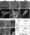
SPK1 and the WAVE complex gene SRA1 function in a common pathway that controls polarized growth. (A) sra1 and spk1 trichomes have a cell-swelling component to their phenotype. SEM of young wild-type (Col), sra1, and spk1 trichomes. (Scale bar: 20 μm.) (B) Actin filament networks are disorganized in the core cytoplasm of growing spk1 and sra1 trichomes. Actin filaments in whole mounted Col, sra1, and spk1 trichomes were visualized with Alexa 488-phalloidin as described in ref. . Confocal images are maximum projections of the entire cell. Arrows indicate developing branch regions that normally contain aligned core actin bundles. (Scale bar: 10 μm.) (C and D) sra1 enhances the cell swelling of the reduced branching mutant glabra3. (C) SEM of the mature trichomes: gl3, gl3 treated with Lat B, and gl3 sra1. (D) Quantitative analysis of single and double mutant trichome phenotypes. Cell length was plotted against swelling factor. Swelling factor is measured from orthogonal views and is a dimensionless value based on the perimeter/area ratio of an equilateral triangle (see Materials and Methods). (E and F) spk1 and spk1 sra1 trichomes have indistinguishable swelling phenotypes. (E) SEM of the mature trichomes: spk1, spk1 treated with Lat B, and spk1 sra1. (F) Quantitation of the cell swelling and cell length phenotypes of spk1 and spk1 sra1 as described in D. (Scale bar: 50 μm.)
Fig. 4.
Fig. 4.
The WAVE complex protein SRA1 is a ROP effector, and endogenous SPK1 and WAVE complex proteins form complexes. (A) SRA1 is an effector for a subset of ROPs. Soluble, purified GST–ROPs were pulled down with bead-bound HIS–SRA1 (Upper) and MBP–RIC1 (Lower) and probed with a α-ROP antibody. The input fraction and the bead-bound fractions from each ROP binding reaction are labeled. The nucleotide status of ROP in each reaction is indicated. (B) Endogenous SPK1 associates with the WAVE complex subunit NAP1 in cells. coIP of SPK1 was performed as described in Fig. 2A. (Upper) Lanes 1 and 4, 10% of input protein extracts from Col or spk1-1 plants, respectively; lanes 2, 5 and 3, 6 are coIP fractions using either α-SPK1N antibody or preimmune serum, respectively. The blot was probed with α-SPK1N (Upper) or α-NAP1 (Lower) antibody. (C) Endogenous SPK1 complexes are smaller when isolated in the sra1 and nap1 backgrounds. Crude extracts containing solubilized SPK1 were analyzed by size-exclusion chromatography. Column fractions were isolated from Col (Top), sra1 (Middle), and nap1 (Bottom). Apparent masses for column fractions are labeled. Vo, void volume. All fractions were probed with the α-SPK1N antibody.

Similar articles

Cited by

References

    1. Molendijk AJ, Ruperti B, Palme K. Small GTPases in vesicle trafficking. Curr Opin Plant Biol. 2004;7:694–700. - PubMed
    1. Yang Z, Fu Y. ROP/RAC GTPase signaling. Curr Opin Plant Biol. 2007;10:490–494. - PMC - PubMed
    1. Schmidt A, Hall A. Guanine nucleotide exchange factors for Rho GTPases: Turning on the switch. Genes Dev. 2002;16:1587–1609. - PubMed
    1. Rossman KL, Channing JD, Sondek J. GEF means go: Turning on RHO GTPases with guanine nucleotide-exchange factors. Nat Rev Mol Cell Biol. 2005;6:167–180. - PubMed
    1. Basu D, El-Assal SE, Le J, Mallery EL, Szymanski DB. Interchangeable functions of Arabidopsis PIROGI and the human WAVE complex subunit SRA-1 during leaf epidermal development. Development. 2004;131:4345–4355. - PubMed

Publication types

MeSH terms

Substances