. 2008 Mar 11;105(10):3825-30.
doi: 10.1073/pnas.0709717105. Epub 2008 Feb 28.

Conditional MHC class I ligands and peptide exchange technology for the human MHC gene products HLA-A1, -A3, -A11, and -B7

Affiliations

Conditional MHC class I ligands and peptide exchange technology for the human MHC gene products HLA-A1, -A3, -A11, and -B7

Arnold H Bakker et al. Proc Natl Acad Sci U S A. .

Abstract

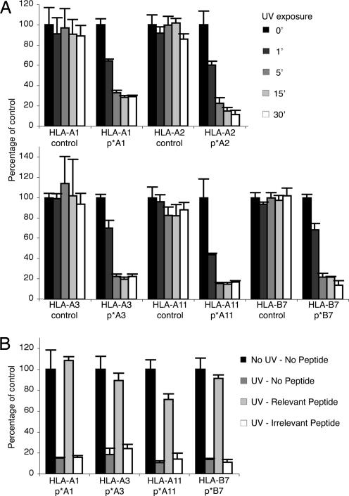
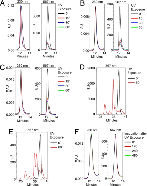
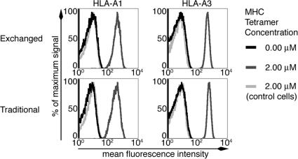
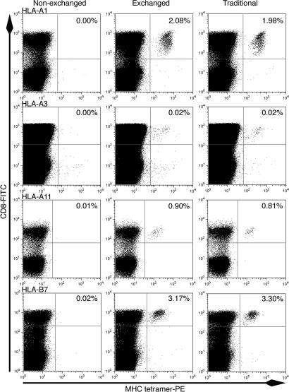
Major histocompatibility complex (MHC) class I multimer technology has become an indispensable immunological assay system to dissect antigen-specific cytotoxic CD8(+) T cell responses by flow cytometry. However, the development of high-throughput assay systems, in which T cell responses against a multitude of epitopes are analyzed, has been precluded by the fact that for each T cell epitope, a separate in vitro MHC refolding reaction is required. We have recently demonstrated that conditional ligands that disintegrate upon exposure to long-wavelength UV light can be designed for the human MHC molecule HLA-A2. To determine whether this peptide-exchange technology can be developed into a generally applicable approach for high throughput MHC based applications we set out to design conditional ligands for the human MHC gene products HLA-A1, -A3, -A11, and -B7. Here, we describe the development and characterization of conditional ligands for this set of human MHC molecules and apply the peptide-exchange technology to identify melanoma-associated peptides that bind to HLA-A3 with high affinity. The conditional ligand technology developed here will allow high-throughput MHC-based analysis of cytotoxic T cell immunity in the vast majority of Western European individuals.


Conflict of interest statement

Conflict of interest statement: The MHC exchange technology described in this manuscript is the subject of a patent application. Based on Netherlands Cancer Institute policy on management of intellectual property, M.T., H.O. and T.N.M.S. would be entitled to a portion of received royalty income in case of future licensing.

Figures

Fig. 1.
Fig. 1.
Characterization of conditional ligands for HLA-A1, -A3, -A11, and -B7. (A) Control and p*-HLA complexes for each complex were exposed to UV for the indicated times, and UV-induced MHC unfolding was measured by ELISA. Control peptides: pA1 VTEHDTLLY, pA2 FLWGPRALV, pA3 RLRAEAQVK, pA11 IVTDFSVIK, and pB7 RPHERNGFTVL. (B) The indicated p*-HLA complexes were exposed to UV light for 0 or 60 min in the presence of no peptide, the HLA-A2 restricted CMV-pp65 epitope NLVPMVATV (irrelevant peptide), or their respective specific ligands, pA1, pA3, pA11, and pB7 (sequences under A) and analyzed by MHC-ELISA. Values indicate means ± SD of triplicates.
Fig. 2.
Fig. 2.
UV-induced peptide cleavage and exchange kinetics. (A) HLA-A2-monomers refolded with the fluorescent UV-sensitive peptide Flp*A2 were treated with UV in the presence of the HLA-A2 ligand NLVPMVATV for different time periods and analyzed by gel-filtration HPLC. Absorption at 230 nm (Left) and fluorescence at 567 nm (Right) was measured. (B and C) HLA B7-monomers refolded with either the fluorescent UV-sensitive peptides AARC(Fl)JTLAM (B) and AARGJTLC(Fl)M (C) were treated with UV in the presence of the HLA-B7 ligand TPRVTGGGAM for different time periods and analyzed as in A. (D) HLA-A2-monomers refolded with the fluorescent UV-sensitive peptide Flp*A2 were either left untreated or exposed to UV for 60 min. Extracted peptides were analyzed by reverse-phase HPLC. Black line, untreated; red line, UV-treated. (E) As in D except that before peptide extration, elution material with the retention time of pMHC molecules was isolated by gel-filtration HPLC. Black line, untreated; red line, UV-treated. (F) p*A2-monomers were treated with UV for 30 min in the presence of 0.5 μM fluorescent FLPSDC(FL)FPSV and 49.5 μM FLPSDCFPSV peptide and kept at temperature for the indicated periods before analysis as in A.
Fig. 3.
Fig. 3.
CD8+ T cell clones specific for HLA-A1 CMV-pp65 (Left) and HLA-A3 EBV-EBNA-3a (Right) were stained with equal amounts of HLA-A1 CMV-pp65 or HLA-A3 EBV-EBNA-3a tetramers, generated via peptide exchange (Exchanged, Upper) or classical refolding (Traditional, Lower). Control cells represent staining with the matched MHC tetramer and cross-sample controls.
Fig. 4.
Fig. 4.
Indicated pMHC complexes were prepared by classical refolding reactions or by 1-h exchange reactions and converted to tetramers. As a negative control, streptavidin-conjugated nonexchanged p*-MHC complexes were used. HLA-typed peripheral blood mononuclear cells were stained with the indicated pMHC tetramers and analyzed by flow cytometry: from top to bottom: HLA-A1 CMV-pp50 (VTEHDTLLY); HLA-A3 EBV-EBNA-3a (RLRAEAQVK); HLA-A11 EBV-EBNA-3b (IVTDFSVIK); HLA-B7 CMV-pp65 (TPRVTGGGAM) tetramers, respectively. For all complexes, stainings were performed at equal concentrations for all three columns. Numbers indicate the percentage of MHC tetramer+ cells of CD8+ cells.

Similar articles

Cited by

References

    1. Altman JD, Moss PA, Goulder PJ, Barouch DH, McHeyzer-Williams MG, Bell JI, McMichael AJ, Davis MM. Science. 1996;274:94–96. - PubMed
    1. Chen DS, Soen Y, Stuge TB, Lee PP, Weber JS, Brown PO, Davis MM. PLoS Med. 2005;2:e265. - PMC - PubMed
    1. Soen Y, Chen DS, Kraft DL, Davis MM, Brown PO. PLoS Biol. 2003;1:E65. - PMC - PubMed
    1. Stone JD, Demkowicz WE, Jr, Stern LJ. Proc Natl Acad Sci USA. 2005;102:3744–3749. - PMC - PubMed
    1. Ljunggren HG, Stam NJ, Ohlen C, Neefjes JJ, Hoglund P, Heemels MT, Bastin J, Schumacher TN, Townsend A, Karre K, et al. Nature. 1990;346:476–480. - PubMed

Publication types

MeSH terms