. 2008 May;26(5):1315-24.
doi: 10.1634/stemcells.2007-0751. Epub 2008 Feb 28.

Evolution of the c-kit-positive cell response to pathological challenge in the myocardium

Affiliations

Evolution of the c-kit-positive cell response to pathological challenge in the myocardium

Jenna Fransioli et al. Stem Cells. 2008 May.

Abstract

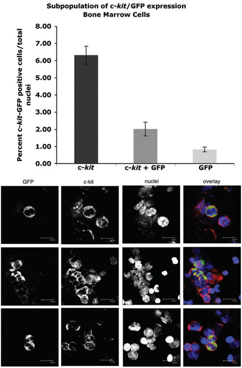
Cumulative evidence indicates that myocardium responds to growth or injury by recruitment of stem and/or progenitor cells that participate in repair and regenerative processes. Unequivocal identification of this population has been hampered by lack of reagents or markers specific to the recruited population, leading to controversies regarding the nature of these cells. Use of a transgenic mouse expressing green fluorescent protein driven by the c-kit promoter allows for unambiguous identification of this cell population. Green fluorescent protein (GFP) driven by the c-kit promoter labels a fraction of the c-kit+ cells recognized by antibody labeling for c-kit protein. Expression of GFP by the c-kit promoter and accumulation of GFP-positive cells in the myocardium is relatively high at birth compared with adult and declines between postnatal weeks 1 and 2, which tracks in parallel with expression of c-kit protein and c-kit-positive cells. Acute cardiomyopathic injury by infarction prompts increased expression of both GFP protein and GFP-labeled cells in the region of infarction relative to remote myocardium. Similar increases were observed for c-kit protein and cells with a slightly earlier onset and decline relative to the GFP signal. Cells coexpressing GFP, c-kit, and cardiogenic markers were apparent at 1-2 weeks postinfarction. Cardiac-resident c-kit+ cell cultures derived from the transgenic line express GFP that is diminished in parallel with c-kit by induction of differentiation. The use of genetically engineered mice validates and extends the concept of c-kit+ cells participating in the response to myocardial injury.


Conflict of interest statement

Disclosure of Potential Conflicts of Interest: The authors indicate no potential conflicts of interest.

Figures

Figure 1
Figure 1
GFP colocalizes with a subpopulation of c-kit-positive bone marrow cells in c-kit-GFP transgenic mice. Top: Histogram representing the number of c-kit-positive, c-kit + GFP-positive, and GFP-positive bone marrow cells (*,p < .05; **, p < .001) (n = 4). Bottom: Confocal scans show representative fields of adult bone marrow cells labeled with antibodies to c-kit (red), GFP (green), and ToPro nuclear stain (blue), as shown in single-channel scans along the left of each color overlay. Abbreviation: GFP, green fluorescent protein.
Figure 2
Figure 2
Developmental expression of c-kit and GFP in mouse myocardium assessed by confocal microscopy in c-kit-GFP transgenic mice. (A): Representative scans of myocardial sections at the time points indicated in upper right corner of each micrograph. Stem cells are indicated (yellow arrows), with c-kit (red), GFP (green), ToPro (magenta), and tropomyosin (red) as shown in single-channel scans along the left of each color overlay. (B): Histogram derived from microscopy shows developmental expression of c-kit+/GFP– (red), c-kit+/GFP+ (yellow), and c-kit–/GFP+ (green) at the time points indicated along the x-axis (n = 3). Abbreviation: GFP, green fluorescent protein.
Figure 3
Figure 3
Developmental expression of c-kit and GFP in mouse myocardium of c-kit-GFP transgenic mice. (A): Immunoblot of whole heart lysates of 2-day-old, 1-week-old, 2-week-old, and 10-week-old mice labeled for c-kit or GFP, with GAPDH used to control for minor variation in sample loading. c-kit+ control is a sample of cultured c-kit+ cells derived from the heart. (B): Quantitation of c-kit and GFP protein levels normalized to those of 2-day old mice. Results are expressed as the mean ± SEM of 2-day old mice. n = 3 for all mice. Abbreviations: GAPDH, glyceraldehyde-3-phosphate dehydrogenase; GFP, green fluorescent protein.
Figure 4
Figure 4
Increases in c-kit+ and GFP+ cells in the myocardium of c-kit-GFP transgenic mice following infarction injury. (A): Immunoblot of c-kit and GFP protein levels in infarct and remote regions of myocardium, with GAPDH as loading control to control for minor variations in protein loading. Positive controls to show antibody recognition include cultured stem cells (+c-kit control) and α-myosin heavy chain driving GFP whole heart lysate (+GFP control). (B): Quantitation of c-kit and GFP protein levels in myocardial lysates of tissue excised from the infarct region 1, 4, 7, 10, and 14 days post-MI relative to sham-operated control tissue (n = 3). (C, D): Evolution of c-kit+ and GFP+ cellular response to MI in c-kit-GFP transgenic myocardium at early (C) or late (D) time points, as indicated in upper right corner of each micrograph. (C): Representative confocal scans of infarcted myocardium labeled at 1 and 4 days post-MI together with sham-operated control, with coincidence of c-kit+ (blue) and GFP (green) shown (yellow arrows), as shown in single-channel scans along the left of each color overlay. Viable myocardium is labeled by sarcomeric tropomyosin (red), whereas nuclei are stained with ToPro dye (magenta). (D): Representative confocal scans of infarcted myocardium labeled at 7, 10, and 14 days post-MI, with coincidence of c-kit+ (red) and GFP (green) shown (yellow arrows), as shown in single-channel scans along the left of each color overlay. Infarct and border zone are labeled as appropriate. Viable myocardium was labeled by sarcomeric tropomyosin (blue), whereas nuclei were stained with ToPro dye (magenta). Changes in color designations between (A) and (B) series scans were made to highlight the relevant cell population in scan sets. Abbreviations: d, day; GAPDH, glyceraldehyde-3-phosphate dehydrogenase; GFP, green fluorescent protein; MI, myocardial infarction.
Figure 5
Figure 5
Colocalization of c-kit+/GATA4+ cells in the infarct region at late stages of time course. (A): Representative confocal scans of infarcted myocardium labeled at 10 days post-MI with coincidence of c-kit+ (red) and GATA4 (green) as indicated (yellow arrows), as shown in single-channel scans along the left of each color overlay. (B): Representative confocal scan of infarcted myocardium labeled at 14 days post-MI, with coincidence of c-kit+ (red) and GATA4 (green) as indicated (yellow arrows). Sarcomeric tropomyosin (magenta) and nuclei (blue) are also shown. Boxed inset regions in the upper right of each panel show higher magnification of the cell from the scan, as indicated. Abbreviations: d, days; MI, myocardial infarction.
Figure 6
Figure 6
Coincidence of GFP+ cells with markers of vascular and endothelial lineages in the c-kit-GFP transgenic mouse. Shown are representative confocal scans of myocardium at 7 (A, B) and 14 (C, D) days postinfarction. Sections were labeled for either α-SMA or von Willebrand's (both shown in yellow) in conjunction with GFP (green), ToPro for nuclei (magenta), and sarcomeric tropomyosin (red). Coincidence of GFP with either α-SMA or von Willebrand's is indicated (arrows in overlay scans; arrowheads in the single-channel scans along the left of each color overlay). Boxed regions (C1, D1) show detail of cells indicated by asterisks in low-power scans. Abbreviations: d, days; GFP, green fluorescent protein; MI, myocardial infarction; α-SMA, α-smooth muscle actin.
Figure 7
Figure 7
Cardiac stem cell (CSC) cultures from hearts of c-kit-GFP transgenic mice show markers of cardiogenic lineage. Undifferentiated CSCs were c-kit+ (A-D), GFP+ (B), nucleostemin+ (C), and Ki67+ (D). Colors for the labels of each protein are shown in the names of each single-channel scan along the left of the corresponding color overlay, with nuclei of cells indicated in blue. Upon induction of differentiation, CSC cultures switched from c-kit+/GFP+ (E) to c-kit–/GFP– (F) and showed increased immunoreactivity for MEF2C in the differentiated (H) versus undifferentiated (G) state. Abbreviation: GFP, green fluorescent protein.

References

    1. Lensch MW, Daheron L, Schlaeger TM. Pluripotent stem cells and their niches. Stem Cell Rev. 2006;2:185–202. - PubMed
    1. Jahagirdar BN, Verfaillie CM. Multipotent adult progenitor cell and stem cell plasticity. Stem Cell Rev. 2005;1:53–59. - PubMed
    1. Beltrami AP, Barlucchi L, Torella D, et al. Adult cardiac stem cells are multipotent and support myocardial regeneration. Cell. 2003;114:763–776. - PubMed
    1. Guan K, Hasenfuss G. Do stem cells in the heart truly differentiate into cardiomyocytes? J Mol Cell Cardiol. 2007;43:377–387. - PubMed
    1. Bearzi C, Rota M, Hosoda T, et al. Human cardiac stem cells. Proc Natl Acad Sci U S A. 2007;104:14068–14073. - PMC - PubMed

Publication types

MeSH terms