. 2008 Apr 25;102(8):914-22.
doi: 10.1161/CIRCRESAHA.107.165530. Epub 2008 Feb 28.

Bone morphogenetic protein-9 is a circulating vascular quiescence factor

Affiliations

Bone morphogenetic protein-9 is a circulating vascular quiescence factor

Laurent David et al. Circ Res. .

Abstract

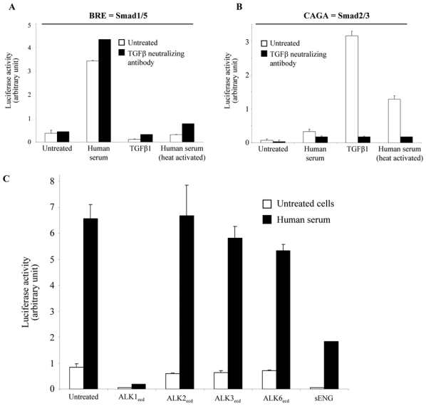
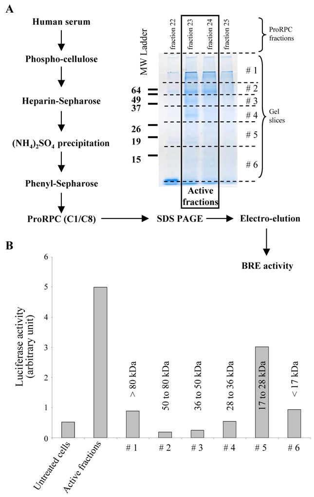
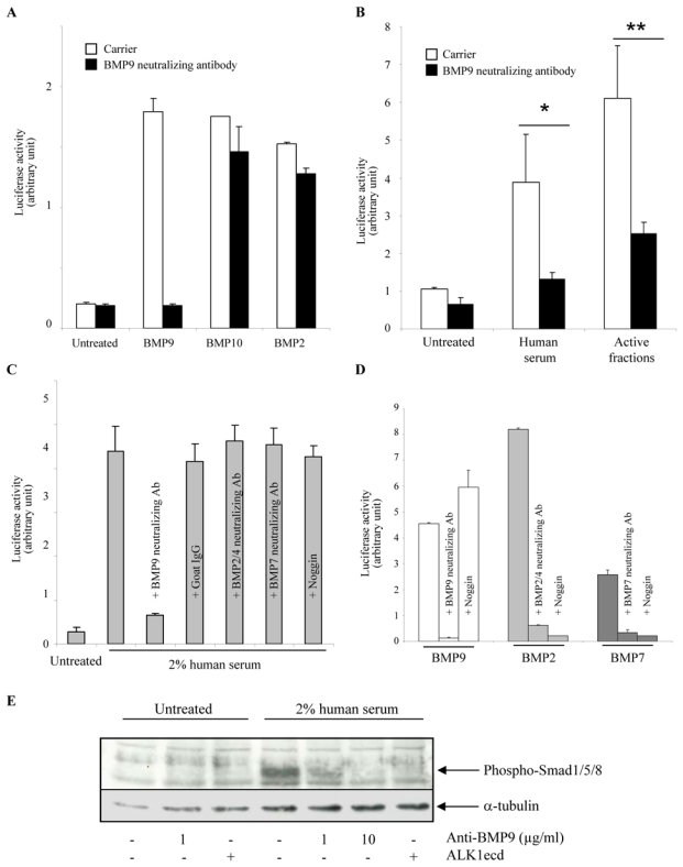
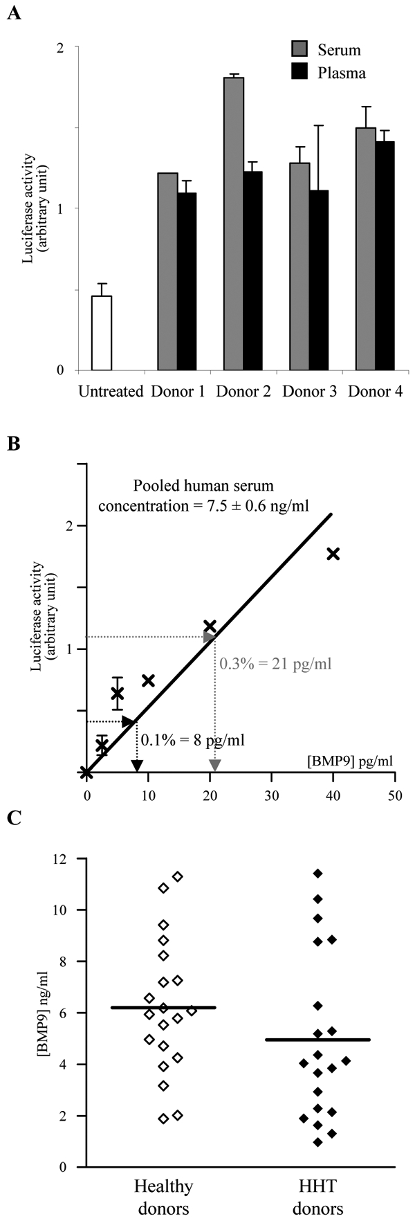
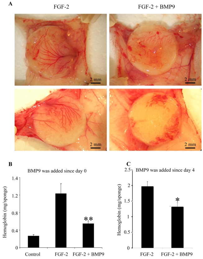
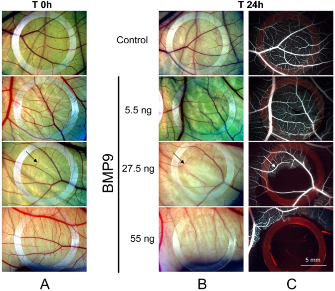
Angiogenesis is a complex process, requiring a finely tuned balance between numerous stimulatory and inhibitory signals. ALK1 (activin receptor like-kinase 1) is an endothelial-specific type 1 receptor of the transforming growth factor-beta receptor family. Heterozygotes with mutations in the ALK1 gene develop hereditary hemorrhagic telangiectasia type 2 (HHT2). Recently, we reported that bone morphogenetic protein (BMP)9 and BMP10 are specific ligands for ALK1 that potently inhibit microvascular endothelial cell migration and growth. These data lead us to suggest that these factors may play a role in the control of vascular quiescence. To test this hypothesis, we checked their presence in human serum. We found that human serum induced Smad1/5 phosphorylation. To identify the active factor, we tested neutralizing antibodies against BMP members and found that only the anti-BMP9 inhibited serum-induced Smad1/5 phosphorylation. The concentration of circulating BMP9 was found to vary between 2 and 12 ng/mL in sera and plasma from healthy humans, a value well above its EC(50) (50 pg/mL). These data indicated that BMP9 is circulating at a biologically active concentration. We then tested the effects of BMP9 in 2 in vivo angiogenic assays. We found that BMP9 strongly inhibited sprouting angiogenesis in the mouse sponge angiogenesis assay and that BMP9 could inhibit blood circulation in the chicken chorioallantoic membrane assay. Taken together, our results demonstrate that BMP9, circulating under a biologically active form, is a potent antiangiogenic factor that is likely to play a physiological role in the control of adult blood vessel quiescence.


Figures

Figure 1
Figure 1. Presence of an ALK1 ligand in human serum that differs from TGFβ
NIH-3T3 cells were transiently transfected with pALK1 and pRL-TK-luc and either pGL3(BRE)-luc (A) or pGL3(CAGA)12-luc (B). Transfected cells were then treated either with human serum (2%), TGFβ1 (0.5 ng/ml) or heat-activated human serum (2%) with or without pan-specific neutralizing TGFβ antibody (1 μg/ml). C: NIH-3T3 cells were transiently transfected with pGL3(BRE)-luc, pALK1 and pRL-TK-luc. Transfected cells were then treated with human serum (2%) in presence or absence of either ALK1ecd, ALK2ecd, ALK3ecd, ALK6ecd or soluble endoglin (200 ng/ml). The luciferase activities were then measured as described in Materials and Methods. Data shown in A, B and C are expressed as mean values ± SD from a representative experiment out of three.
Figure 2
Figure 2. Purification and estimation of the molecular weight of the ALK1 ligand from the human serum
A: Scheme of purification of ALK1 ligand from 250 ml of a pool of human sera. The proteins present in the active fractions (23 and 24) of the Pro-RPC column and the two surrounding fractions (22 and 25), as determined with the BRE reporter gene assay (see Material and Methods), were then separated by 12% SDS-PAGE. After the migration, the gel (fractions 23 and 24) was sliced into 6 parts as indicated by the dotted lines and the proteins were electro-eluted. B: NIH-3T3 cells were transiently transfected with pGL3(BRE)-luc, pALK1 and pRL-TK-luc. Transfected cells were then treated with 100 μl of either the active fractions (fraction 23 and 24) or 100 μl of the proteins eluted from each gel slice. The luciferase activities were then measured as described in Materials and Methods. Data are expressed as mean values ± SD from a representative experiment out of three.
Figure 3
Figure 3. The ALK1 activity of the human serum is due to BMP9
A, B, C and D: NIH-3T3 cells were transiently transfected with pGL3(BRE)-luc, pRL-TK-luc and pALK1. A: Transfected cells were then treated with BMP9 (0.1 ng/ml), or BMP10 (20 ng/ml), or BMP2 (100 ng/ml) in the presence or the absence of a neutralizing BMP9 antibody (1 μg/ml) or an isotype-matched control antibody (1μg/ml). B: Transfected cells were then treated with human serum (1%) or 100 μl of active fraction (fractions 23 and 24 of Fig. 2A). C: Transfected cells were treated with 2% human serum in the presence or the absence of neutralizing antibodies (anti-BMP9 (2 μg/ml), anti-BMP2/4 (10 μg/ml), or anti-BMP7 (10 μg/ml)) or with recombinant noggin (1 μg/ml). D: Transfected cells were treated with either BMP9 (0.05 ng/ml), BMP2 (50 ng/ml) or BMP7 (100 ng/ml) in the presence or the absence of neutralizing antibodies (anti-BMP9 (2 μg/ml), anti-BMP2/4 (10 μg/ml), or anti-BMP7 (10 μg/ml)) or with recombinant noggin (1 μg/ml). The luciferase activities were then measured as described in Materials and Methods. Data shown in A, B, C and D are expressed as mean values ± SD from a representative experiment out of three. E: HMVEC-d were serum-starved for 1 h and were then treated with 2% human serum for 1 h in the presence or absence of neutralizing BMP9 antibody (1 or 10 μg/ml) or ALK1ecd (100 ng/ml). Cell lysates (20 μg proteins) were resolved by 10% SDS-PAGE, and immunoblotted with antibodies against phosphoSmad1/5/8 or against α-tubulin.
Figure 4
Figure 4. Determination of BMP9 concentration in human serum
A: NIH-3T3 cells were transiently transfected with pGL3(BRE)-luc, pRL-TK-luc, pALK1. Transfected cells were treated with 0.5% of human serum or plasma of 4 different healthy donors. B: linear regression for the determination of BMP9 serum concentration. NIH-3T3 cells were transiently transfected with pGL3(BRE)-luc, pRL-TK-luc, pALK1. Transfected cells were then treated with 0.1 or 0.3% of a pool of human sera. The luciferase activities were then measured as described in Materials and Methods. Data shown in A and B are expressed as mean values ± SD from a representative experiment out of three. C: BMP9 serum levels measured in 20 patients with HHT and 20 healthy donors. The line indicates the mean value. The difference was not statistically significant.
Figure 5
Figure 5. Effect of BMP9 on angiogenesis in the mice sponge assay
A and B: Bal-C mice received a subcutaneous cellulose sponge treated with FGF-2 (200 ng) and/or BMP9 (20 ng) under the dorsal skin. Injections in the sponge of FGF-2 and/or BMP9 diluted in PBS were performed on day 1 and day 2 and a last injection was performed on day 4 with BMP9 alone. C: Bal-C mice received a subcutaneous cellulose sponge treated with FGF-2 (200 ng) diluted in PBS under the dorsal skin. Injections of FGF-2 were performed on day 1 and day 2. BMP9 (20 ng) or PBS were injected on day 4, 5 and 6. Animals were sacrificed on day 7 and the sponges were photographed (A). Hemoglobin content was measured in 1 ml of RIP A buffer extract of the sponge and adjacent vascular network (B and C). B: Data are expressed as mean values ± SEM from a representative experiment (five mice in each groups) out of three. C: Data are expressed as mean values ± SEM of two experiments (nine mice in each groups). (* p < 0.05; ** p < 0.01).
Figure 6
Figure 6. Effect of BMP9 on vessel formation in the chick chorioallantoic membrane assay
On day 9, the CAM received either 25 μl of BMP9 (5.5 ng, 27.5 ng, 55 ng or 550 ng) or vehicle (Control). The photographs shown were taken before (T 0h) and after treatment (T 24h) and are representative of the results obtained in an additional five eggs per group. Low magnification pictures of CAMs at T 0h(A) and T 24h (B); C: 24 h after treatment, FITC dextran was injected in the CAM vessels, fluorescent images. Arrow indicates a vessel that is not affected by BMP9 treatment; arrowhead indicates a vessel that cannot be seen after BMP9 treatment.

References

    1. Xiao YT, Xiang LX, Shao JZ. Bone morphogenetic protein. Biochem Biophys Res Commun. 2007;362:550–553. - PubMed
    1. Miller AF, Harvey SA, Thies RS, Olson MS. Bone morphogenetic protein-9. An autocrine/paracrine cytokine in the liver. J Biol Chem. 2000;275:17937–17945. - PubMed
    1. Lopez-Coviella I, Berse B, Thies RS, Blusztajn JK. Upregulation of acetylcholine synthesis by bone morphogenetic protein 9 in a murine septal cell line. J Physiol Paris. 2002;96:53–59. - PubMed
    1. Truksa J, Peng H, Lee P, Beutler E. Bone morphogenetic proteins 2, 4, and 9 stimulate murine hepcidin 1 expression independently of Hfe, transferrin receptor 2 (Tfr2), and IL-6. Proc Natl Acad Sci U S A. 2006;103:10289–10293. - PMC - PubMed
    1. Johnson DW, Berg JN, Baldwin MA, Gallione CJ, Marondel I, Yoon SJ, Stenzel TT, Speer M, Pericak-Vance MA, Diamond A, Guttmacher AE, Jackson CE, Attisano L, Kucherlapati R, Porteous ME, Marchuk DA. Mutations in the activin receptor-like kinase 1 gene in hereditary haemorrhagic telangiectasia type 2. Nat Genet. 1996;13:189–195. - PubMed

Publication types

MeSH terms