. 2008 Mar;154(Pt 3):865-877.
doi: 10.1099/mic.0.2007/013292-0.

Wall teichoic acids are dispensable for anchoring the PNAG exopolysaccharide to the Staphylococcus aureus cell surface

Affiliations

Wall teichoic acids are dispensable for anchoring the PNAG exopolysaccharide to the Staphylococcus aureus cell surface

Marta Vergara-Irigaray et al. Microbiology (Reading). 2008 Mar.

Abstract

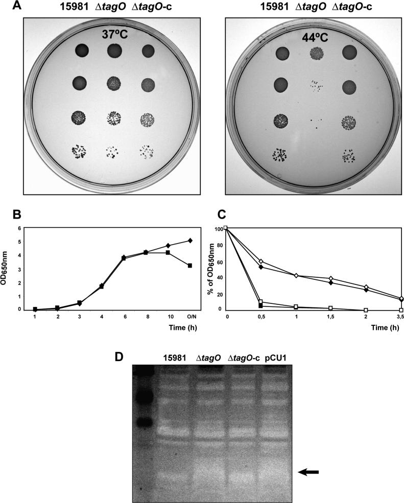
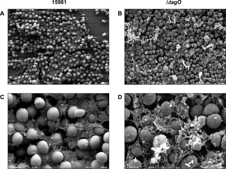
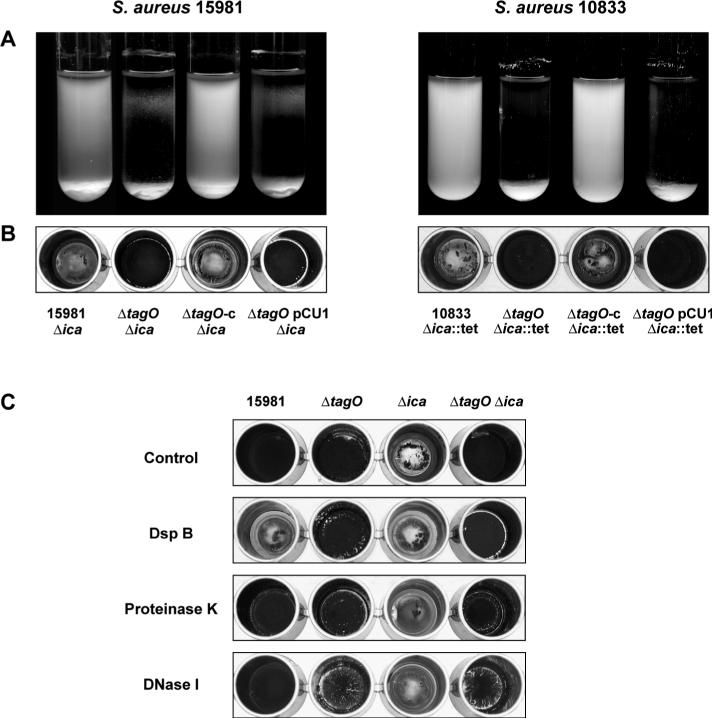
Biofilm formation in Staphylococcus aureus is usually associated with the production of the poly-N-acetylglucosamine (PNAG) exopolysaccharide, synthesized by proteins encoded by the icaADBC operon. PNAG is a linear beta-(1-6)-linked N-acetylglucosaminoglycan that has to be partially deacetylated and consequently positively charged in order to be associated with bacterial cell surfaces. Here, we investigated whether attachment of PNAG to bacterial surfaces is mediated by ionic interactions with the negative charge of wall teichoic acids (WTAs), which represent the most abundant polyanions of the Gram-positive bacterial envelope. We generated WTA-deficient mutants by in-frame deletion of the tagO gene in two genetically unrelated S. aureus strains. The DeltatagO mutants were more sensitive to high temperatures, showed a higher degree of cell aggregation, had reduced initial adherence to abiotic surfaces and had a reduced capacity to form biofilms under both steady-state and flow conditions. However, the levels as well as the strength of the PNAG interaction with the bacterial cell surface were similar between DeltatagO mutants and their corresponding wild-type strains. Furthermore, double DeltatagO DeltaicaADBC mutants displayed a similar aggregative phenotype to that of single DeltatagO mutants, indicating that PNAG is not responsible for the aggregative behaviour observed in DeltatagO mutants. Overall, the absence of WTAs in S. aureus had little effect on PNAG production or anchoring to the cell surface, but did affect the biofilm-forming capacity, cell aggregative behaviour and the temperature sensitivity/stability of S. aureus.


Figures

Figure 1
Figure 1. The absence of WTAs in S. aureus impairs growth under high temperatures and induces autolysis
A) Effect of high temperatures on growth. S. aureus 15981, 15981 ΔtagO and complemented mutant 15981 ΔtagO-c, were grown exponentially in TSB-gluc at 37°C to an OD650nm of 0.2. The cultures were then serially diluted (10−1, 10−2, 10−3, and 10−4 fold) and 10 μl of each dilution were spotted onto TSA plates. The plates were then incubated at 37°C or 44°C overnight. B) Growth curve of the wild type strain 15981 (&#x25C6) and ΔtagO mutant (■). C) Triton X-100 induced autolysis assay. The autolysis of mid-exponential phase cultures of wild type strain 15981 (&#x25C6), 15981 ΔtagO mutant (■), complemented mutant 15981 ΔtagO-c (&#x25C7) and control strain 15981 ΔtagO carrying pCU1 plasmid (□) was determined at 37°C in the presence of 0.05 % Triton X-100, by measuring the decrease in the optical density at 650 nm upon exposure to the detergent. D) Zymogram analysis. Cell extracts were applied to a SDS polyacrylamide gel containing S. aureus RN4220 cells and stained with 1% methylene blue. Areas of murein hydrolase activity are indicated by clear zones. Lane 1, molecular size marker; lane 2, parental strain 15981; lane 3, ΔtagO mutant; lane 4, complemented mutant ΔtagO-c and lane 5, control ΔtagO strain carrying pCU1 plasmid. The arrow highlights the zone of major differences.
Figure 2
Figure 2. Biofilm formation and aggregative phenotype of two genetically unrelated S. aureus strains and their respective ΔtagO mutants
A) Biofilm forming capacity on glass surfaces of overnight cultures incubated in TSB-gluc medium, with shaking, at 37°C of S. aureus wild type strains 15981 (strong biofilm former) and 10833 (weak biofilm former), their isogenic ΔtagO mutants, the complemented ΔtagO mutants and the control ΔtagO strains carrying empty pCU1 plasmid. B) Biofilm forming capacity on microtiter plates after 24 h of static incubation at 37°C in TSB-gluc medium. C) Biofilm formation on microtiter plates after 24 h of static incubation at 37°C in TSB-gluc medium. The pellicle formed on the bottom of the plates were air-dried before staining. D) Comparison of the primary attachment ability of S. aureus 15981, its ΔtagO mutant, the complemented mutant and the control ΔtagO strain carrying pCU1 plasmid. Data represent the means of at least 9 counts from three independent experiments. The vertical line at the top of the each bar represents the standard deviation. Very significant differences were detected between wild-type strain and ΔtagO mutant (ns, not significant versus wild type; *** P < 0.001 versus wild type; one-way ANOVA test with Tukey's pairwise comparisons)
Figure 3
Figure 3. Scanning electron microscopy analysis of ΔtagO mutant
Scanning electron microscopy images of S. aureus 15981 (A and C) and its ΔtagO mutant (B and D) at magnifications of × 5000 and × 20000.
Figure 4
Figure 4. Biofilm formation of S. aureus 15981 and ΔtagO mutant under continuous flow conditions
A) Biofilm development in microfermentors of wild type S. aureus strain 15981 and ΔtagO mutant grown under continuous-flow conditions with TSB-gluc after 24 h at 37°C. The microfermentors (upper panels) contain the glass slides where bacteria form the biofilm (lower panels). The results of a representative experiment are shown. B) Quantification of the biofilm mass adherent to the glass slides. The cells were removed from the glass slides into 20 ml of TSB-gluc by vortexing, and the OD of the resulting solutions was measured at 650 nm. Significant differences were detected between the wild-type and the ΔtagO mutant (n = 4; P < 0.05, Mann-Whitney U test)
Figure 5
Figure 5. Dot-blot analysis of PNAG accumulation
A) Dot-blot analysis of surface localized PNAG levels of S. aureus wild type strain 15981, its ΔtagO mutant, the complemented mutant and the control ΔtagO strain carrying the empty pCU1 plasmid. Cell surface extracts of overnight cultures were treated with proteinase K and dilutions of the samples (1/50, 1/75 and 1/100) were spotted onto nitrocellulose membranes. PNAG production was detected with rabbit antiserum raised to deacetylated PNAG conjugated to diphtheria toxoid. As negative controls, a Δica mutant unable to produce PNAG was used. B) Dot-blot analysis of PNAG cell surface levels of overnight cultures using different extraction conditions ranging from harsher to milder solvents (Urea 4M > NaCl 1M > NaCl 0.8M) and (EDTA, 100°C > EDTA, room temperature > water, 100°C > water, room temperature).
Figure 6
Figure 6. Immunoelectron microscopy
Immunoelectron microscopy images probed first with rabbit antiserum raised to deacetylated PNAG conjugated to diphtheria toxoid and then with gold-labeled donkey-anti-rabbit IgG. A-B) S. aureus 15981, C-D) S. aureus 15981 ΔtagO mutant, E) S. aureus 15981 Δica mutant and F) S. aureus 15981 ΔicaB mutant.
Figure 7
Figure 7. Effect of ΔicaADBC deletion and biofilm detachment treatments
A) Biofilm forming capacity on glass surfaces of overnight cultures incubated in TSB-gluc medium, with shaking, at 37°C of S. aureus Δica strains, their ΔtagO mutants and the double mutants carrying the plasmid pCU1 containing the tagO gene under the control of its own promoter or alone. B) Biofilm formation on microtiter plates after 24 h of static incubation at 37°C in TSB-gluc medium. The biofilms formed on the bottom of the plates were air-dried before staining. C) Susceptibility of ΔtagO biofilms to enzymatic treatments. Biofilms of 15981, ΔtagO, Δica and ΔtagO Δica strains were grown in TSB-gluc for 24 h and treated during 2 h at 37 °C with Dispersin B or proteinase K. DNase I was added at time of inoculation and biofilms were incubated in TSB-gluc for 24 h. The bacteria that remained attached to the surface were air-dried and stained with crystal violet.

References

    1. Aly R, Shinefield HR, Litz C, Maibach HI. Role of teichoic acid in the binding of Staphylococcus aureus to nasal epithelial cells. J Infect Dis. 1980;141:463–465. - PubMed
    1. Arnaud M, Chastanet A, Débarbouillé M. A new vector for efficient allelic replacement in naturally non transformable low GC% Gram-positive bacteria. Appl Environ Microbiol. 2004;70:6887–6891. - PMC - PubMed
    1. Augustin J, Rosenstein R, Wieland B, Schneider U, Schnell N, Engelke G, Entian KD, Gotz F. Genetic analysis of epidermin biosynthetic genes and epidermin-negative mutants of Staphylococcus epidermidis. Eur J Biochem. 1992;204:1149–1154. - PubMed
    1. Baddiley J, Buchanan JG, Hardy FE, Martin RO, Rajbhandary UL, Sanderson AR. The structure of the ribitol teichoic acid of Staphylococcus aureus H. Biochimica et biophysica acta. 1961;52:406–407. - PubMed
    1. Baddiley J. Teichoic acids in bacterial coaggregation. Microbiology. 2000;146(Pt 6):1257–1258. - PubMed

Publication types

MeSH terms

LinkOut - more resources